A black scab on a wound is often eschar, dry, dead tissue, rather than a normal crust. Whether it needs medical care depends on its stability, location, blood flow, and signs of infection, not color alone.
You notice it while changing a dressing: a dark patch covers the wound, and it's hard to tell whether healing is going normally. A regular scab can look dark, too, but black tissue may also hide a deeper wound or a circulation problem. The important questions are what the tissue is, whether it's stable, and what signs mean you should seek care promptly.
## Table of Contents - What a Black Scab on a Wound Usually Means - A calm way to judge what you're seeing - How Eschar Differs from a Normal Healing Scab - Clues you can observe without touching - Why Wound Tissue Turns Black - Why color alone cannot grade severity - Stable Eschar and Warning Signs to Watch For - When not to wait - How Clinicians Evaluate and Treat Black Wound Tissue - What happens at an assessment - Choosing a removal method - When a Black Scab Signals a Stalled Wound - Signs a wound may need a closer look - Key Takeaways and Your Next Step
What a Black Scab on a Wound Usually Means
A dark covering may be an ordinary crust of dried blood and wound fluid, or it may be eschar, a layer of dead, nonviable tissue. Those can look similar at a glance, but they don't mean the same thing for the wound. A crust can protect healing skin; eschar may cover tissue that needs clinical assessment.
For example, a small, dry crust over a scrape may be part of routine healing. A thick black area on a heel or foot, especially if it's changing, painful, draining, or surrounded by redness, deserves more attention. The appearance alone can't show how deep the injury goes or whether blood reaches the area adequately.
A calm way to judge what you're seeing
Start with the details around the black area, not just its color. Consider where it is, whether it feels dry or wet, whether it's firmly attached or lifting, and whether there's new drainage, odor, pain, redness, or warmth. Diabetes, pressure on the area, and known circulation problems also matter.
A black covering doesn't automatically mean an emergency, and it doesn't automatically mean the wound is healing safely. Stable tissue in a particular location may be left intact while a clinician checks circulation. Unstable tissue or signs of infection call for prompt assessment. The sections below explain how to distinguish a normal crust from eschar and why “don't pick it” is sound advice, but not a complete treatment plan.
How Eschar Differs from a Normal Healing Scab
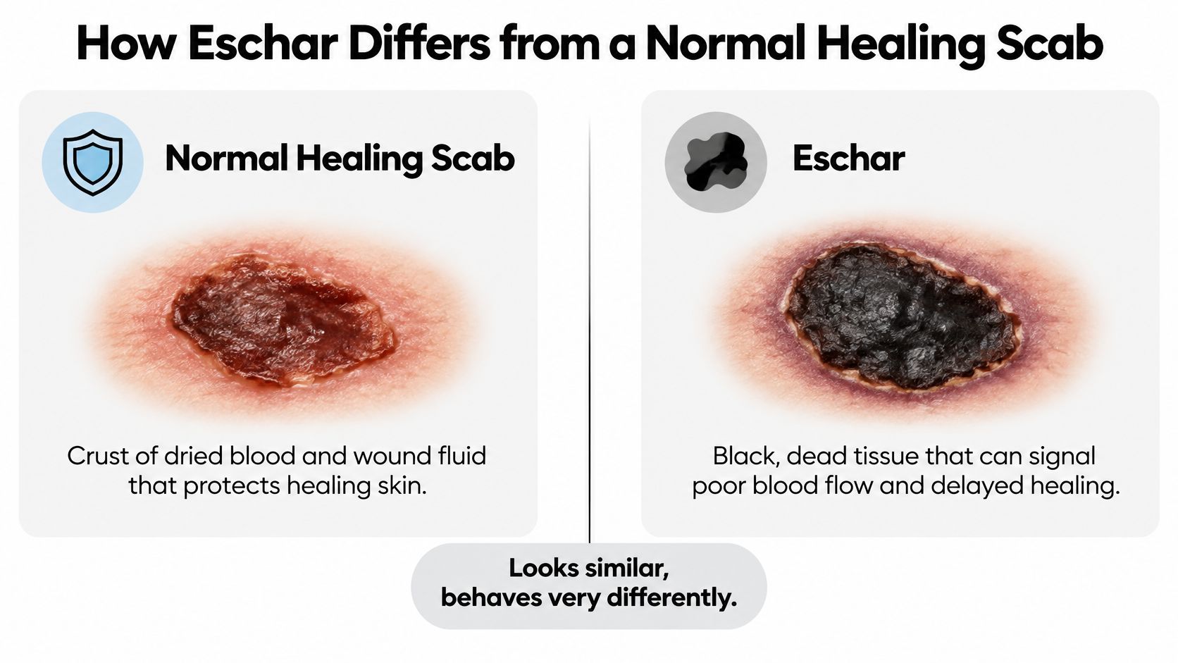
Start with what the covering is made of. A normal scab is dried blood and wound fluid that forms a temporary lid while new skin grows underneath. It often looks dark red or brown, feels crusty, and loosens at the edges as repair progresses. Peeling it early can reopen the surface.
Eschar is dead tissue, not dried fluid. It tends to be thicker and may look black, dark brown, or tan. The texture is often dry, hard, or leathery, though infected tissue can become soft or boggy. Because it is part of the wound rather than a layer on top, it can block oxygen, trap bacteria, and keep the wound bed hidden.
Clues you can observe without touching
Look at the margin and the trend. A healing scab usually gets smaller, drier, and less tender as days pass. Eschar is more likely to stay firmly stuck, cover a depression, sit over a pressure point, or come with swelling, drainage, odor, warmth, or pain. Location raises the stakes: black tissue over a heel, toe, sacrum, or diabetic foot makes circulation and pressure central questions.
Color alone cannot settle it. Some safe scabs look nearly black, and some serious wounds are partly yellow, red, or tan. The safer home judgment uses trend and context, then a clinician checks blood flow, depth, infection risk, and pressure before deciding whether black tissue should be left stable or removed.
Practical rule: A crust can be left alone while it lifts naturally. Suspected eschar needs a wound and circulation assessment, especially on the heel or foot, before anyone decides removal is safe.
Why Wound Tissue Turns Black
A black area usually means one of two things is happening on the surface: blood and fluid have dried into a crust, or underlying tissue has been injured enough to die. The color is a clue, not a diagnosis.
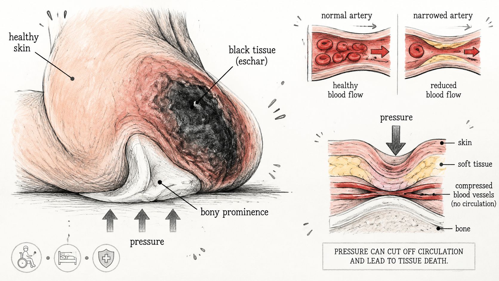
Blood supply is the common thread. Skin stays alive because arteries deliver oxygen and nutrients while veins carry waste away. Sustained pressure over a heel, tailbone, or ankle can squeeze those vessels closed; shear can twist them; diabetes, peripheral artery disease, swelling, infection, trauma, or surgery can reduce flow further. When flow stays too low for too long, cells die, the wound bed breaks down, and necrotic tissue can darken from tan to brown to black.
Why color alone cannot grade severity
A thin scab over a shallow scrape is dried protein and blood sitting above living skin. True eschar behaves more like a leathered lid of dead tissue attached to the wound, and it can hide a crater that reaches fat, tendon, muscle, or bone. As noted earlier, that is why pressure injuries covered by eschar are treated as unstageable until a clinician can see the base.
This is also why “never remove black tissue” is incomplete advice. Before deciding whether dead tissue should come off, clinicians check circulation and look for infection. Removing eschar without enough arterial flow can leave a larger wound that cannot close, while infected or unstable tissue may need prompt care. A stable, dry heel eschar can be the exception, which is why the blood-flow check comes before the removal decision.
Stable Eschar and Warning Signs to Watch For
Some black tissue can be left in place while a clinician assesses the wound. A dry, firmly attached, intact heel eschar without redness or fluctuance is a special case. It may act as a natural cover, and wound-care guidance generally advises against routinely removing it. Hospital wound-care guidance on stable heel eschar
That doesn't mean every dry black area is safe to watch without guidance. If the tissue becomes wet, loose, draining, or soft, or if the surrounding skin changes, the situation may need a different plan. The table compares features that can help you describe what you see to a clinician.
| Characteristic | Likely Stable Eschar | Signs That Need Prompt Assessment |
|---|---|---|
| Surface and attachment | Dry, intact, firmly adherent | Wet, soft, loose, cracked, or separating |
| Surrounding skin | No new redness or swelling | Spreading redness, warmth, or swelling |
| Drainage and odor | No drainage or foul smell | Drainage, pus, or foul odor |
| Pain and texture | No increasing pain or bogginess | Rising pain or soft, boggy tissue |
| Location and health factors | Stable heel area under clinical observation | Necrotic heel or foot wound with diabetes, or a wound with circulation concerns |
| General symptoms | No fever or feeling acutely unwell | Fever or other signs of systemic illness |
When not to wait
A black area that is expanding, wet, lifting, foul-smelling, or increasingly painful should be assessed promptly. The same applies to spreading redness or warmth, swelling, purulent drainage, or fever. These changes can indicate unstable necrosis or infection and shouldn't be treated as routine scabbing.
Diabetes changes the stakes for wounds on the heel or foot. Guidance calls for urgent specialist referral when a person with diabetes has a necrotic wound in these locations. If you're unsure how quickly a wound needs attention, CWS's wound escalation pathway describes an option for surgical wound evaluation.
How Clinicians Evaluate and Treat Black Wound Tissue
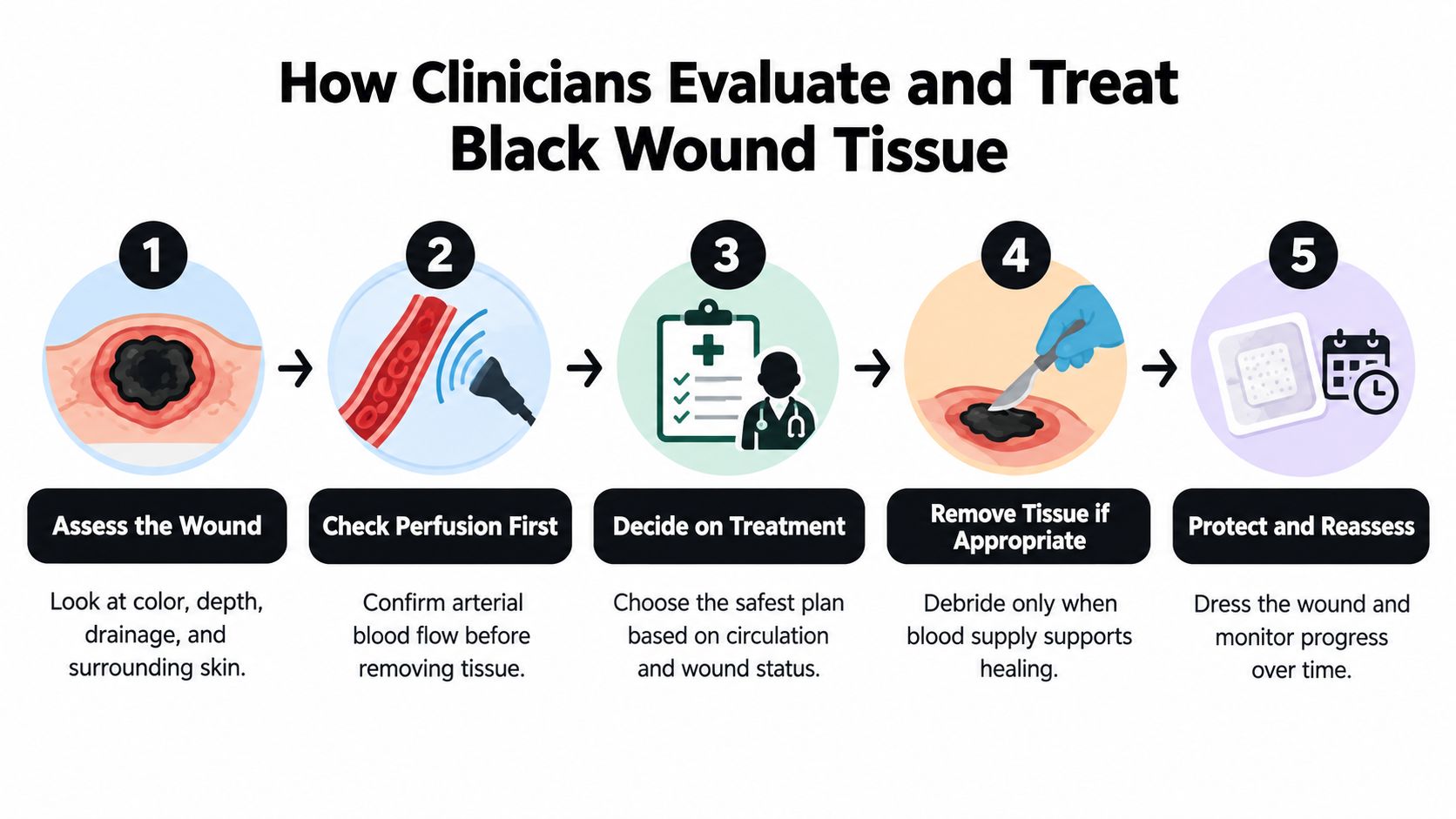
Removing black tissue without first checking the blood supply works like rewiring a house before confirming power reaches it. The first question a clinician asks is whether enough arterial blood reaches the wound, a check called perfusion assessment. This matters most for wounds on the foot, heel, or lower leg, and for people with diabetes, vascular or kidney disease, a smoking history, or absent pulses. Debriding a wound with poor arterial flow can leave an open area the body cannot repair. Guidance on perfusion assessment before debridement
What happens at an assessment
A clinician records the wound's location, dimensions, and tissue type, along with drainage, odor, pain, redness, and fluctuance, meaning a soft, fluid-filled feel under the skin. Circulation, pressure on the area, blood sugar control, and infection signs all factor in. The goal is to answer three questions: should this tissue stay, does the wound need imaging or lab work, and which removal method fits if one is needed.
The perfusion answer can override everything else. A dry, stable heel eschar may be deliberately left intact while blood flow is assessed, which is why evaluation comes before removal rather than after it.
Choosing a removal method
When removal is appropriate, the method depends on depth, blood supply, infection status, bleeding risk, and the person's overall health. Options include sharp or surgical debridement, enzymatic agents that dissolve dead tissue, and autolytic debridement, where the body's own moisture loosens it. A method that suits one wound can harm another. Wound-management guidance on debridement choices
Don't cut, scrape, soak, or apply over-the-counter products to remove a black scab yourself. It may cover deeper tissue or an area with poor circulation, and uncontrolled removal can cause bleeding or worsen the injury.
Wound Services treats complex, stalled, and surgically appropriate wounds, meeting patients virtually or in person. A wound surgeon reviews your history, images, and the wound itself by video, usually within days. Its advanced wound care service and surgical wound evaluation are available when a wound needs this level of assessment.
When a Black Scab Signals a Stalled Wound
Black tissue that persists, returns, or keeps covering the wound can mean the wound isn't progressing as expected. Removing visible dead tissue, when appropriate, doesn't fix the reason it formed. Pressure, reduced blood supply, infection, trauma, surgery, or systemic disease may still be affecting healing. The cause needs attention alongside any decision about the tissue itself. Guidance on managing lower-leg eschar and escalating care
Treatment that relies on the body gradually breaking down tissue can take longer and isn't suitable for every wound. It may be inappropriate for dry, hard eschar when arterial insufficiency is present, and it isn't preferred in some infected wounds. The cited wound-care guideline recommends referral when lower-leg eschar is present and escalation if an initial autolytic approach hasn't worked within one week. That guidance underscores why persistent black tissue merits reassessment rather than repeated home treatment.
Signs a wound may need a closer look
Wound Services lists indicators that a wound may deserve surgical evaluation, including necrotic tissue, no measurable size reduction, persistent undermining, infected tissue or recurrent cellulitis, exposed tendon or bone, persistent slough, recurrent breakdown, and minimal improvement despite appropriate evidence-based care. Learn more about stalled wounds and evaluation
For a virtual consultation, a wound surgeon reviews the patient's history, images, and wound by video, usually within days. A surgical wound-care nurse then validates findings in person. When an intervention is appropriate, the surgeon who evaluated the wound is the one who operates and follows healing afterward.
That process doesn't mean every wound needs surgery. It gives the care team a way to reconsider the wound's cause and whether the current plan is working, while preserving conservative treatment when that's appropriate.
Key Takeaways and Your Next Step
A black covering may be a normal crust or eschar, and color alone can't tell you which. Pay attention to the wound's location, whether the tissue is dry and firmly attached or wet and separating, and whether pain, redness, drainage, odor, swelling, or fever is developing. A stable heel eschar may be left intact under clinical guidance, but changing or infected-looking tissue needs prompt assessment.
Don't scrape or cut the area at home. For a useful record, take dated photographs, note the wound's size, and write down any changes in drainage, smell, pain, surrounding redness, or general symptoms. Share that information with a clinician, especially if the wound persists, recurs, or is on the foot or heel in someone with diabetes.
Wound Services reports that approximately 18,521 wounds have improved or healed through its care, and that its average time to healing is less than 21 days. Individual results vary with wound type, health history, and care plan. Persistent black tissue isn't a reason to assume the worst, but it is a reason to find out what's preventing the wound from healing.
---
If you're concerned about a black scab that's changing, not improving, or covering a complex wound, Wound Services offers virtual and in-person wound evaluation, with surgery considered only when clinically appropriate. Visit Comprehensive Wound Services to explore an evaluation and discuss the next step for your wound.