
You're halfway through a dressing change when the gauze lifts and the base looks pink, shiny, and a little wet. Someone nearby says it finally looks like healing. That red, moist layer is often wound granulation tissue, and it can mean repair is underway. It can also sit there for weeks while the wound barely shrinks. What follows explains what that tissue is, how long it should last, and which changes should send you to a clinician instead of another week of the same dressing.
## Table of Contents - Why a Red, Moist Wound Bed Matters - What the color can and can't prove - What Granulation Tissue Is Made Of - The jelly that holds the work - What to write down after each dressing change - Where Granulation Fits in the Healing Timeline - Day ten beside day thirty - Why Granulation Is Not the Finish Line - A patch on a shirt - Healthy Granulation Versus Hypergranulation - Overgrowth is not a lab result - Signs That Warrant Clinical Review - What the bed should feel like - Choosing the Right Next Step for a Stalled Wound - Three realistic paths
Why a Red, Moist Wound Bed Matters
A caregiver feels relief at that color, and the relief makes sense. Pink or red tissue usually means blood is reaching the bed, which black or thick yellow tissue can't offer. The error is treating color as proof the wound is closing. A bed can look busy for weeks and barely shrink. Tissue can also look encouraging while it has grown too high for skin cells to cross.
Wound granulation tissue is the temporary, highly vascular fill of the proliferative phase, after bleeding stops and early inflammation settles. It isn't new skin. It fills the defect, gives migrating skin cells a surface, and helps form a barrier while repair continues. Later, scar is supposed to replace it. If that fill lingers, overgrows, or never shows up, both the wound and the person's overall health need another look.
What the color can and can't prove
- Shade: Bright red and moist usually fits active repair. Pale, gray, or dusky tissue is a different finding.
- Level: Tissue inside the wound bed is expected. Tissue mounded above the skin edge is not the same thing.
- Change: A bed that looks identical across several dressing changes, with no smaller outline, isn't quiet success.
Practical rule: Photograph the wound in the same light and from the same distance once a week. Compare the outline and the edges, not only how red the center looks.
If the bed is red, level with or slightly below the skin, moist without soaking the dressing, and the outline is slowly shrinking, stay with the plan your clinician already set and keep those photos. If the tissue rises, bleeds when you barely touch it, smells worse, drains more, hurts more, or the wound reopens, call that week. Another seven days of the same dressing won't answer those signs. From here, the parts of the tissue, the normal clock, the line between healthy fill and overgrowth, and the call-a-clinician list all connect to that same decision.
What Granulation Tissue Is Made Of
Think of the wound as a building that lost a wall. Granulation tissue is the scaffold, not the finished rooms. Workers and supplies move through it. Once the permanent structure is up, that scaffold is supposed to come down.
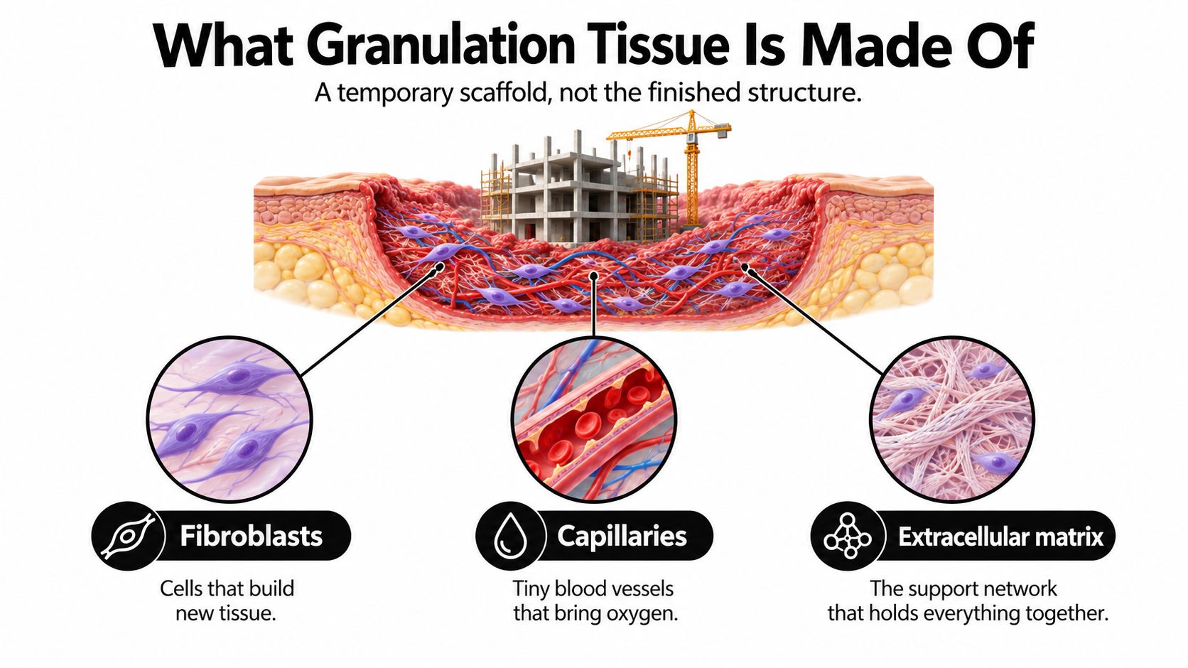
Three kinds of cells do most of the visible work.
Fibroblasts are the builders. They move in and lay down new collagen and other matrix materials. Endothelial cells line the new capillaries and drive angiogenesis, new vessel growth, under signals that include vascular endothelial growth factor, fibroblast growth factor, platelet-derived growth factor, and transforming growth factors, as a granulation tissue review describes. Keratinocytes are the skin cells that later crawl across the surface. Macrophages and other inflammatory cells clear debris and help keep the repair signal going. No single cell type is "the tissue" by itself.
The jelly that holds the work
Around those cells sits a loose extracellular matrix of collagen, fibronectin, and hyaluronic acid. Early collagen is a softer form that can still be rearranged. The matrix is a temporary jelly cells can migrate through. It isn't the tougher, quieter collagen of an old scar.
Thin-walled capillaries run through that jelly. Small bumps on the surface often match vessel loops pushing upward. Plasma leaks from those thin walls, which is why a healthy bed looks moist as well as red. The tissue is metabolically busy. It is spending oxygen and nutrients, not sitting there as a cosmetic patch. Calling it new skin skips that job.
What to write down after each dressing change
"Granulation present" is a weak note. A useful record includes:
- Fill: About how much of the base is this red tissue, as a share of the bed.
- Color and moisture: Red and moist, or pale, gray, dry, or soupy.
- Firmness, odor, and drainage: Resilient versus fragile, and whether smell or fluid is rising.
- Size: Length, width, and depth, plus whether the edge is advancing.
That list is the biology turned into a habit. You're tracking a scaffold that should change, not admiring a color.
Where Granulation Fits in the Healing Timeline
Healing behaves more like a relay than a single sprint. One phase hands the wound to the next. If a runner holds the baton too long, the race stalls even though people are still on the track.
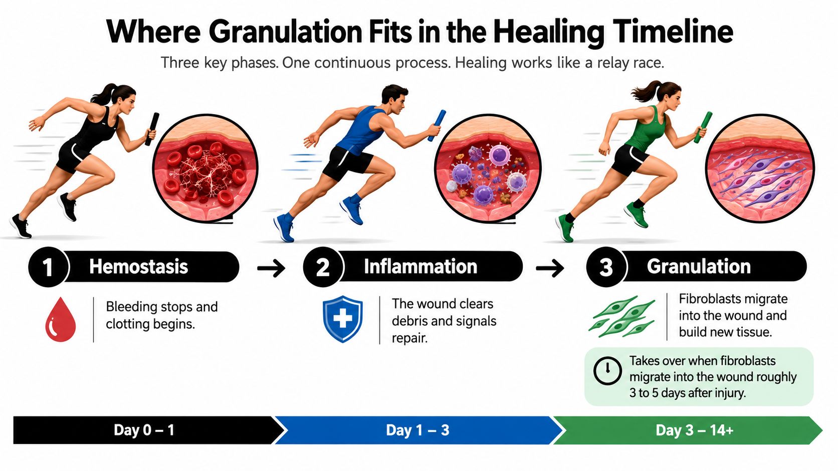
Hemostasis starts the race. Bleeding slows and a clot forms, over minutes to hours. Inflammation clears debris and signals repair. Fibroblasts then migrate into the wound about 2 to 3 days after injury, which is when granulation takes the baton. In an uncomplicated skin wound, that proliferative phase may continue for up to 3 weeks. By about days 5 to 7, fibroblasts have begun depositing new collagen and glycosaminoglycans. Those ranges are laid out in a clinical review of wound healing phases.
Day ten beside day thirty
Around day ten, an uncomplicated wound is often still in that fill-and-rebuild window. A red, moist bed with early edge movement can fit the clock. The tissue is doing its job, and it is still temporary.
By about day thirty, the handoff should be underway. Remodeling typically begins around week 3 and can continue for up to 12 months. The red fill is an intermediate milestone, not the end of repair. A wound that still looks like a fresh construction site at five or six weeks, with little change in size, has missed the expected handoff.
A short video walks through the same sequence if a picture helps more than a calendar.
A wound can be on schedule at day ten and off schedule at day forty with the same red color. The clock is part of the exam.
Why Granulation Is Not the Finish Line
A filled or even closed wound invites a second mistake. People stop protecting it because the surface looks done. The biology isn't done.
Granulation tissue is structurally immature. Published descriptions put its collagen at about 30% to 40% type III collagen, a softer reticular form that acts as an early scaffold. During remodeling, that type III collagen is gradually replaced by stronger type I fibrillar collagen. Net collagen production stays active for roughly 4 to 5 weeks, and the broader shift from type III to type I can run for about a year. Tensile strength is reported to peak at about 80% of uninjured skin around 3 months after injury, according to collagen remodeling data in the wound-healing literature. Repaired skin doesn't fully regain its original strength.
A patch on a shirt
Sew a patch onto torn fabric and the hole looks closed the same day. Pull on the seam that afternoon and the stitches give. Skin is similar. The surface can look continuous while collagen underneath is still being swapped and rewoven. Apparent closure isn't maturity.
A wound that reopens isn't always a minor scrape that "didn't stick." Recurrent breakdown can reflect incomplete remodeling, collagen that hasn't matured, or a biological problem that was never corrected.
- Keep the load low: Follow the offloading, footwear, or pressure plan well after the surface looks quiet.
- Keep the dressing plan: Don't switch to a dry gauze "because it's closed" unless your clinician says the wound is ready.
Teams who need a structured plan for that longer phase can look at advanced wound care instead of assuming a closed surface can carry a normal load.
Healthy Granulation Versus Hypergranulation
The finding that fools people most often is success that has grown too far. Hypergranulation is granulation tissue that rises above the wound edge. It may look lobulated or shiny, it often bleeds easily, and it can physically block the skin cells that need to cross the wound. Image-analysis research notes that this overgrowth is frequently mistaken for healthy granulation, especially early on.
Picture a rim that looks pink and hopeful. The center, though, sits proud of the skin line, glossy, and it bleeds when gauze brushes it. Covering that mound, or trying to stimulate more tissue, doesn't remove the block. Skin cells can't climb a hill that already stands above the shore they're trying to reach.
Overgrowth is not a lab result
Friable or exuberant tissue isn't automatic proof of bacterial infection. Wound literature treats hypergranulation, recurrent breakdown, epithelial bridging, increased exudate, and delayed healing as possible subtle signs of local infection or biofilm. Those problems generally lack a definitive lab test. Clinicians usually identify them at the bedside, and experience varies. That's a reason to escalate, not a reason to start leftover antibiotics at home.
| Feature | Healthy granulation | Hypergranulation or possible infection |
|---|---|---|
| Height | Fills the bed without rising past the skin edge | Mounds above the edge and can block skin cells |
| Surface | Red, moist, slightly pebbled, resilient | Often shiny, lobulated, or overly fragile |
| Bleeding | Limited, not set off by light contact | Bleeds easily with gentle touch |
| Edges | Skin advances and the outline shrinks | Bridging, stalled edges, or recurrent breakdown |
| Drainage and odor | Controlled moisture, stable smell | Increasing drainage, worse odor, or new pain |
| Home decision | Continue the set plan and keep weekly photos | Ask for a clinician's exam rather than another week alone |
If the red tissue is higher than the skin around it, treat that height as a question for a clinician, not as extra healing.
Signs That Warrant Clinical Review
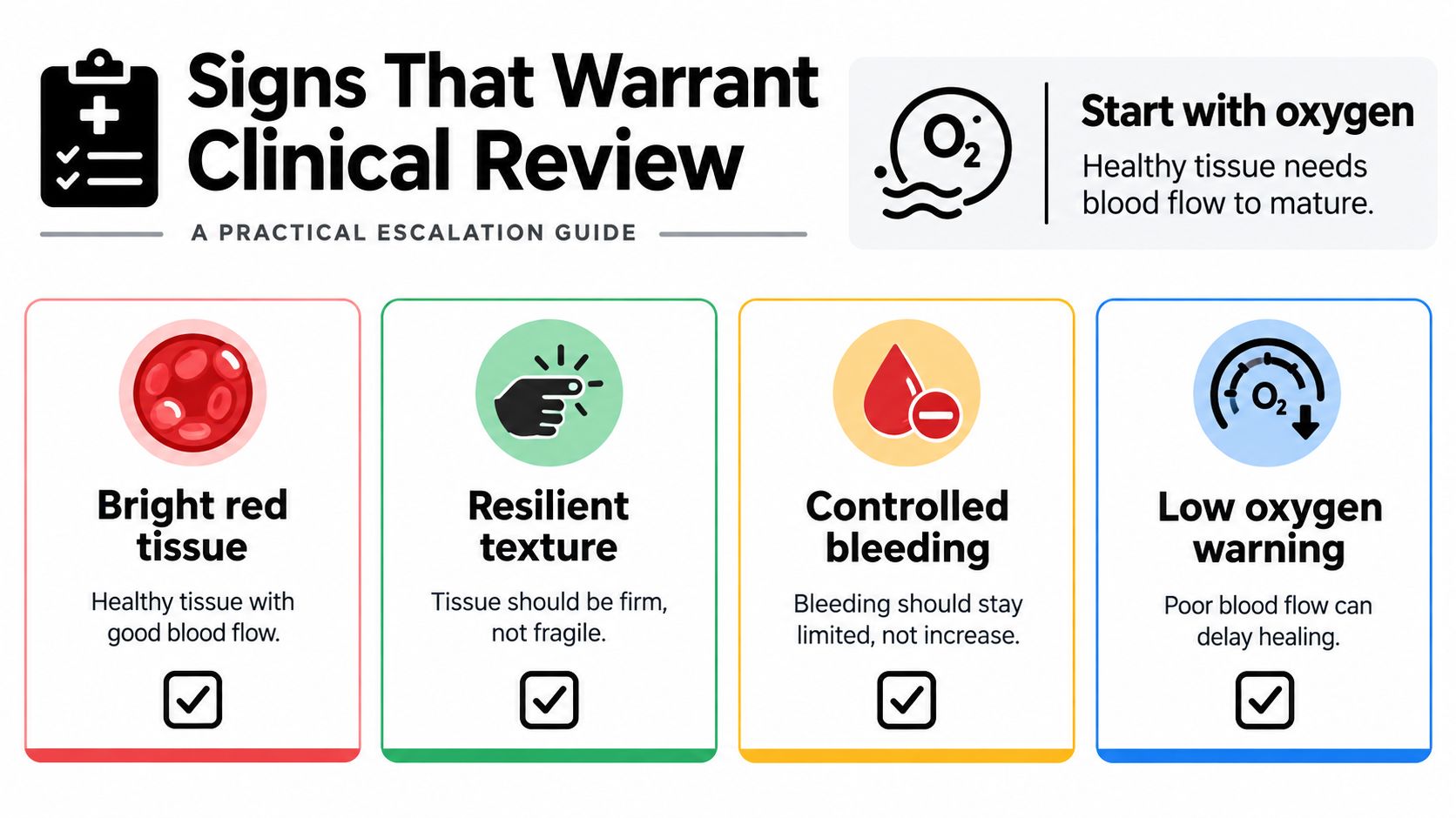
Oxygen is the quiet requirement behind a good-looking bed. It supports vessel growth, fibroblast work, bacterial defense, epithelialization, and collagen maturation. Hydroxylation of proline and lysine during collagen synthesis depends on oxygen. A wound can show living granulation and still stall when perfusion is poor, or when edema, pressure, smoking, vascular disease, anemia, or infection limits delivery. That link is described in work on oxygen and collagen maturation.
What the bed should feel like
Bright red, resilient tissue with controlled drainage generally means vascular repair is active. Dusky, pale, gray, excessively fragile, or rapidly bleeding tissue deserves a check for poor perfusion, ongoing inflammation, trauma, or overgrowth. Raised tissue, covered above, can also block skin migration and shouldn't be dressed as if it were healthy fill.
Call for professional review, rather than another home-only week, when you see:
- Bleeding with gentle contact: New vessels can ooze, but bleeding that starts with light touch and keeps returning needs an exam.
- Tissue above the skin edge: Height that blocks the rim is a mechanical problem.
- Worse odor or drainage: A new smell or a wetter dressing can mean bioburden or inflammation, even without fever.
- More pain, reopening, or a stalled outline: Increasing pain, recurrent breakdown, or no progress in closure is the point to stop waiting.
Our escalation pathway is built for that handoff. We look for eight indicators that a wound may deserve surgical evaluation: no measurable size reduction, necrotic or dead tissue, persistent undermining, infected tissue or recurrent cellulitis, exposed tendon or bone, persistent slough tissue, recurrent wound breakdown, and minimal improvement despite appropriate evidence-based care. Share your photos and dates with the current team the same week if any of those show up beside the color changes above. An evaluation doesn't mean an operation. It means perfusion, pressure, infection, and tissue quality get a plan that can change.
Choosing the Right Next Step for a Stalled Wound
A stronger dressing isn't the only lever when the outline stops shrinking, and it isn't always the right one. A 2025 systematic review of five randomized studies involving 236 patients found enzymatic debridement produced higher granulation and epithelialization rates than autolytic debridement, with complete healing in 65% versus 50% of cases, while still calling for more research before firm guidelines. A 2025 plasma summary reported healing rates of 53.3% with weekly treatment and 61.5% with twice-weekly treatment, versus 25.0% in controls, and noted that protocols and patient selection still matter. Both sets of findings sit in this 2025 evidence summary. More tissue growth, by itself, isn't a sufficient endpoint.
Three realistic paths
- Keep monitoring with the current team when drainage is controlled and measurements show steady shrinkage. Weekly photos and the dressing plan you already have are the right tools.
- Ask that clinician to reassess when progress slows but the tissue is still level and resilient. Pressure, edema, glucose, nutrition, dead tissue, and bioburden can block skin from crossing even when the bed looks red. Sometimes the next step is a cleaner bed, not a different foam.
- Request a specialist review, virtual or in person, when the wound stays stalled, keeps reopening, or shows features that may need a procedure. A wound surgeon reviews the history, the wound, imaging, and the treatment response virtually. A surgical wound care nurse then validates the wound in person so the findings match what was seen on screen. The surgeon who evaluates the wound is the surgeon who operates if intervention is appropriate, and who follows the healing afterward. How that triage works is outlined in our note on when a wound stops progressing.
This week, gather dates, photos, dressing names, and any change in pain, drainage, or size. Send that packet to the clinician already involved. If the measurements aren't moving, ask for specialty review rather than another month of the same routine.
---
Wound Services evaluates complex, stalled, and possibly surgical wounds, with attention to whether granulation is progressing or only looking red. A surgeon can review your history and images virtually, and in-person validation follows when the findings need a closer look. If your wound isn't shrinking, visit Comprehensive Wound Services and ask about a review this week.