
What if a breast wound keeps getting new dressings, but nobody has established why the skin broke down? Breast skin ulcers are a visible finding, not a diagnosis. Infection, cancer, inflammatory disease, treatment-related injury, and other conditions can damage the skin, yet they don't all respond to the same care.
That distinction matters because a wound can look infected without infection being the main cause. Careful assessment should relieve symptoms while identifying the disease underneath, without assuming that every ulcer is cancer or that every open wound needs surgery.
## Table of Contents - Possible Causes of Breast Skin Ulcers at a Glance - How Common Breast Skin Ulcers Are and Who Gets Them - What these estimates mean - The burden goes beyond wound size - How Clinicians Assess a Breast Skin Ulcer - The assessment sequence - Why biopsy can change the plan - When a Breast Ulcer Needs Urgent Specialist Review - Act on these warning signs - Escalate when the explanation no longer fits - A stalled wound also deserves review - Postoperative Breast Breakdown That Is Not Simple Infection - Patterns that should change the discussion - Review the diagnosis before repeating the procedure - Caring for a Breast Ulcer While the Cause Is Unclear - Avoid making a fragile wound worse - Make the care record useful - Confirm the Next Step With Your Care Team - Prepare for a useful consultation
Possible Causes of Breast Skin Ulcers at a Glance
| Cause | Typical clues | Usual first step |
|---|---|---|
| Malignant tumor invasion | Crater-like ulcer or cauliflower-like nodule, bleeding, odor, sometimes a breast mass | Skin biopsy and histopathology |
| Infection such as mastitis or abscess | Fever, spreading redness, swelling, purulent drainage | Clinical evaluation; cultures interpreted alongside examination findings |
| Inflammatory breast cancer | Breast-wide redness or swelling, orange-peel texture, nipple changes | Imaging and biopsy rather than treatment as simple infection |
| Radiation injury | Ulceration within a previously irradiated area, slow healing | Wound assessment and biopsy if persistent or atypical |
| Cutaneous angiosarcoma | Ulcerated skin lesion that can overlap with other skin changes | Skin biopsy and histopathology |
| Postoperative dehiscence | A surgical incision opens, sometimes with drainage | Wound review; consider inflammatory disease if pain is disproportionate |
| Pyoderma gangrenosum | Painful, rapidly progressing ulcer with undermined purple borders | Biopsy planning and multidisciplinary review before more surgery |
| Trauma or factitial injury | Irregular edges, injury history, or repeated picking | Detailed history and exclusion of other causes |
| Medication effects | Thinning skin, stretch marks, or exposed fat after steroid-related skin damage | Medication review with the prescriber |
| Fungal infection | Rash or skin breakdown that needs testing to establish a fungal cause | Skin sampling for fungal testing |
| Inflammatory disease resembling hidradenitis | Recurrent deep nodules and draining tunnels in skin folds | Dermatology review and biopsy when indicated |
The clues are questions to investigate, not diagnostic rules. A draining postoperative wound could represent infection, an opening incision, or an inflammatory disorder. A history of radiation makes treatment-related injury relevant, but it doesn't establish the explanation by itself.
Some entries also describe uncommon but important possibilities. For example, a clinicopathologic study of cutaneous angiosarcoma associated ulceration with poorer overall survival, reinforcing why an unexplained ulcerated skin lesion needs tissue diagnosis rather than assumptions based on appearance.
Use this table as a map of questions to bring to a clinician, not a tool for choosing treatment yourself.
An ulcer means that skin has broken down into an open sore. The task is to determine what caused that breakdown, whether a second problem such as infection has developed, and which interventions are safe.
How Common Breast Skin Ulcers Are and Who Gets Them
A review of fungating and ulcerating breast cancer cites estimates that 5–10% of patients with cancer develop fungating wounds, while approximately 2–5% of patients with locally advanced breast cancer develop a fungating breast wound. These figures concern cancer-related wounds, not every possible cause of breast skin ulceration.
The breast is a common location among malignant fungating wounds, but its share differs between settings. A 2025 review of malignant fungating wounds reported the breast as the most common anatomical site, accounting for 49% of cases. Published estimates shouldn't be treated as a single universal rate.
What these estimates mean
Cancer-related breast ulcers are uncommon relative to all breast cancers and are concentrated among advanced, biologically aggressive, or delayed-presenting disease. Tumor cells can invade the skin and disrupt tissue, producing an ulcer or a raised, fragile wound.
Reliable population counts are difficult because cancer registries don't consistently record fungating wounds. Differences in access to diagnosis, disease stage at presentation, and clinical reporting also affect estimates.
The evidence has limits: a 2024 systematic review of fungating and ulcerating breast cancers included 79 studies, of which 62 were case reports and 7 were case series. That makes the published literature useful for understanding clinical problems, but less reliable for predicting an individual person's course.
The burden goes beyond wound size
Pain, drainage, odor, bleeding, and restricted movement can interfere with sleep, clothing, personal care, and relationships. People may feel embarrassed or avoid seeking help, even though these symptoms deserve treatment without judgment.
Wound care still matters when closure isn't currently feasible. Better symptom control can make everyday activities more manageable while the oncology team considers treatment.
For someone without a known cancer diagnosis, these estimates don't establish the likelihood that a new ulcer is malignant. A new breast ulcer needs evaluation because several different diseases can produce the same visible result.
How Clinicians Assess a Breast Skin Ulcer
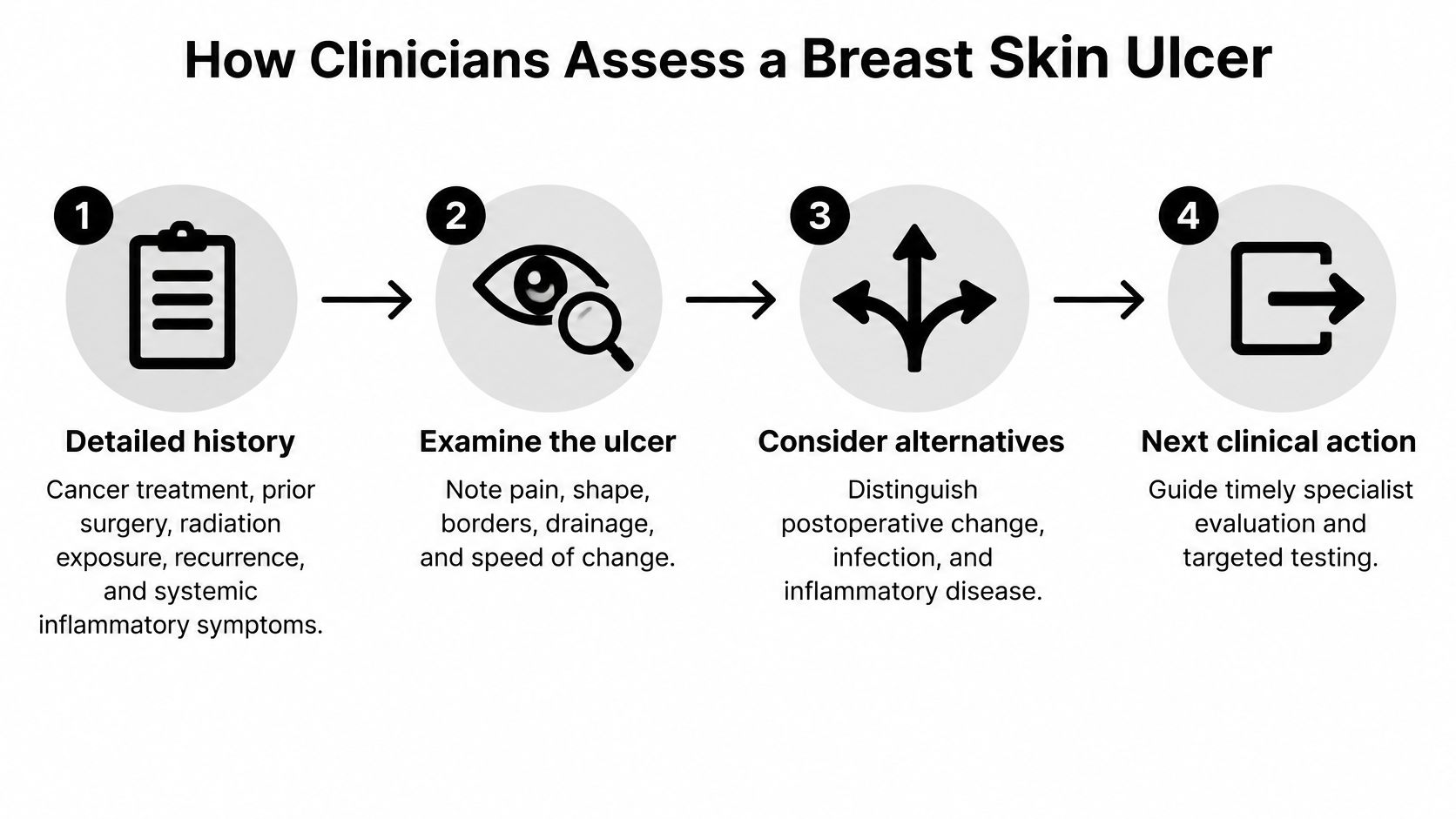
A useful assessment follows a sequence: establish the history, describe the wound, investigate likely causes, and measure the response to treatment. The narrative review of breast ulceration in Advances in Skin & Wound Care supports combining detailed clinical history, reproducible wound documentation, and histopathologic sampling in most cases, particularly persistent or atypical ulcers.
The assessment sequence
- Establish the history. The clinician asks about cancer treatment, previous breast or underarm surgery, radiation exposure, earlier episodes, medications, and inflammatory symptoms elsewhere in the body. When the ulcer started, what preceded it, and how it changed can help separate otherwise similar-looking conditions.
- Examine and measure the wound. Documentation includes its location, type, length, width, depth, and any undermining, where tissue loss extends beneath the visible skin edge. The clinician also records dead tissue and slough, the material that can cover part of the wound bed.
- Describe symptoms and surrounding skin. Drainage amount and character, bleeding, odor, pain, and the condition of the surrounding skin all belong in the record. These observations help the team choose protection and symptom management without relying only on a photograph.
- Evaluate possible secondary infection. Fever, expanding redness, swelling, increasing pain, and pus-like drainage require clinical assessment. A positive culture alone doesn't establish infection, because clinicians must interpret the result alongside the examination and symptoms.
- Plan tissue sampling when indicated. Persistent, recurrent, rapidly progressing, unusual, treatment-resistant ulcers, or ulcers associated with a breast mass, warrant particular attention to biopsy. Histopathology means examining sampled tissue under a microscope to distinguish malignant infiltration from inflammatory disease, radiation injury, and other causes.
- Compare progress with the baseline. Repeat measurements and symptom records show whether the current plan is helping. A wound that looks cleaner but continues enlarging needs a different discussion from one that is becoming smaller and less painful.
Why biopsy can change the plan
A biopsy isn't a final stamp on a diagnosis that everyone already suspects. It can redirect treatment entirely, especially when a presumed infection or ordinary wound has behaved unexpectedly.
For example, a clinician may need to determine whether tissue breakdown reflects tumor involvement or an inflammatory process before deciding whether tissue removal is appropriate. The safest next intervention depends on the cause, not just the amount of damaged skin.
Bring dated photographs, a measurement log if available, the names of dressings and medicines tried, and a symptom diary. Don't probe the ulcer yourself to measure depth or undermine its edges; leave that assessment to the treating team.
When a Breast Ulcer Needs Urgent Specialist Review
Some changes make waiting for a routine appointment unsafe. A clinical review of malignant cutaneous wound management identifies persistent bleeding, exposed deep structures, rapidly increasing necrosis, uncontrolled pain, fever, and spreading redness as reasons for urgent specialist evaluation.
Act on these warning signs
- Heavy or persistent bleeding: Apply firm pressure with a clean dressing while seeking care. Heavy bleeding, bleeding that won't stop, or bleeding accompanied by weakness or faintness needs emergency assessment.
- Fever with spreading redness: Seek same-day emergency care rather than waiting for another dressing appointment.
- Rapidly increasing tissue damage: Expanding areas of dead tissue or visible deep structures need urgent review.
- Uncontrolled pain: Severe pain that isn't controlled by the prescribed plan warrants prompt reassessment.
These findings don't identify the underlying disease on their own. They indicate that the person may need immediate bleeding control, infection assessment, pain treatment, or protection of deeper tissues.
Escalate when the explanation no longer fits
Other changes may be less dramatic but still call for diagnostic escalation: rapid enlargement, a new breast mass, widespread breast swelling, nipple inversion or discharge, or swelling in the armpit. Orange-peel skin texture, called peau d'orange, is another finding to report.
The Royal Australian College of General Practitioners guide to breast skin changes advises reconsidering presumed mastitis when it hasn't improved within about one week of antibiotic treatment. Don't wait for that interval if symptoms are worsening or emergency warning signs develop.
A clinician may arrange mammography, ultrasound, and biopsy even when the lesion looks infected. The possibilities include inflammatory breast cancer, invasive malignancy, Paget disease affecting the nipple area, inflammatory skin disease, fungal infection, and radiation-related injury.
Practical rule: When treatment isn't producing the expected response, ask whether the diagnosis needs to change before simply repeating the treatment.
Say redness was initially treated as mastitis, but the breast becomes more swollen and the nipple changes despite antibiotics. The next step is reassessment of the breast condition, not an indefinite sequence of antibiotic refills or dressing changes.
A stalled wound also deserves review
Our escalation pathway identifies eight findings that may justify surgical wound evaluation: no measurable size reduction, necrotic tissue, persistent undermining, infected tissue or recurrent cellulitis, exposed tendon or bone, persistent slough, recurrent breakdown, and minimal improvement despite appropriate evidence-based care.
An evaluation does not automatically mean surgery. For a breast ulcer, the team must first consider whether the proposed intervention fits the diagnosis and whether oncology or dermatology input is needed.
Wound management and cancer exclusion can proceed together. Taking an ulcer seriously doesn't mean assuming the worst; it means avoiding preventable delay while treating the symptoms already affecting the patient.
Postoperative Breast Breakdown That Is Not Simple Infection
A 2025 case series of breast pyoderma gangrenosum described 10 women, with diagnostic evolution ranging from 4 days to 7 months; six patients with postsurgical disease experienced diagnostic delays of 3–7 months because clinicians didn't initially suspect the condition. The lesson isn't that every postoperative ulcer is inflammatory, but that an unexpected course should challenge the original explanation.
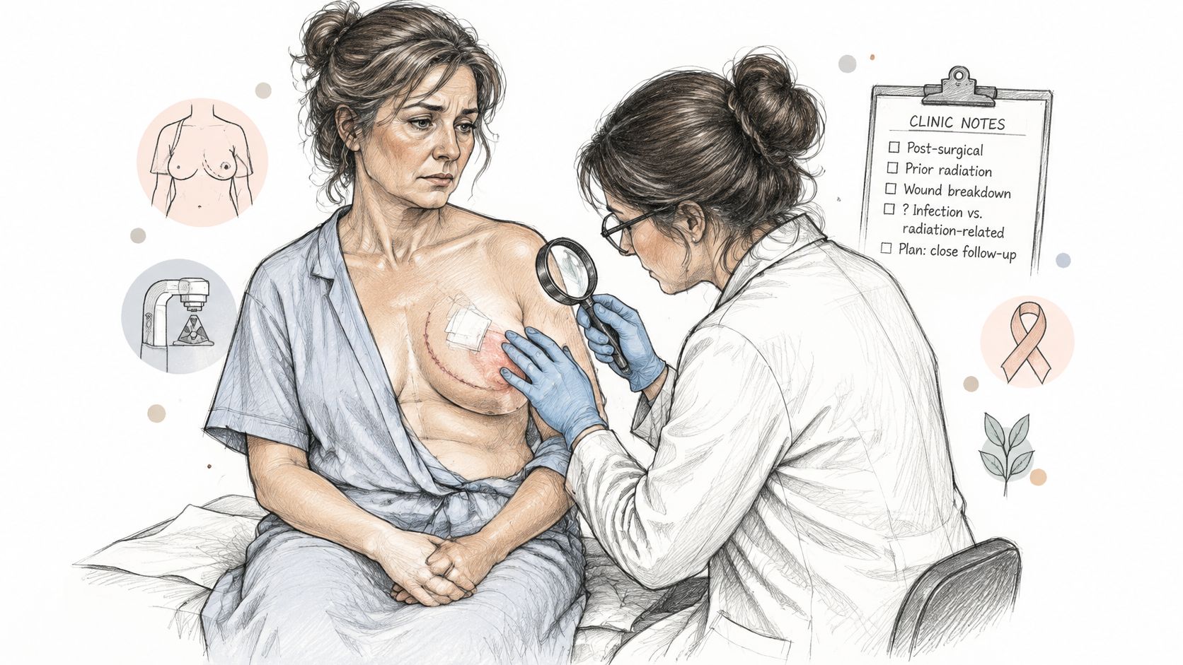
Pyoderma gangrenosum is an inflammatory ulcerative disorder, not a wound infection. In susceptible people, tissue trauma can provoke further ulceration, a response called pathergy.
That creates a difficult clinical trap. A wound worsens, clinicians remove more damaged tissue, and the resulting trauma can worsen the inflammatory lesion rather than resolve it.
Patterns that should change the discussion
- Pain out of proportion: The severity seems greater than the visible wound would suggest.
- Rapid progression: Necrosis or ulceration expands unexpectedly.
- Unusual borders: The edge is purple, undermined, or particularly inflamed.
- Deterioration after procedures: New injury appears or the ulcer expands after intervention.
- An inconsistent infection story: Cultures are sterile or repeatedly fail to explain the course, and antimicrobial treatment doesn't produce the expected improvement.
None of these findings proves pyoderma gangrenosum. Together, however, they support multidisciplinary review involving breast surgery, wound care, dermatology, pathology, and infectious-disease expertise when appropriate.
Review the diagnosis before repeating the procedure
Ordinary wound separation and infection still require appropriate wound care, and an abscess may need drainage. The concern arises when the wound doesn't follow the expected course despite a reasonable treatment plan.
Radiation injury is a separate possibility that also needs its own assessment. A wound in treated skin shouldn't automatically be assigned the same explanation or procedure as a routine incision breakdown.
For a surgical wound evaluation, bring the operative history, culture results, photographs taken before and after procedures, and pathology reports. These help the reviewing clinician distinguish persistent disease from a complication of treatment.
Don't refuse urgently needed treatment based on a suspected diagnosis alone. Instead, ask the team to explain how the findings support the planned intervention and whether biopsy needs special planning.
Before another procedure, ask whether this painful wound has been evaluated for an inflammatory disorder.
Caring for a Breast Ulcer While the Cause Is Unclear
Symptom relief shouldn't wait for every diagnostic result, but home care shouldn't replace assessment. Until the cause is established, keep self-care focused on basic protection and the treating clinician's instructions.
The following measures are discussion points, not a complete home-treatment protocol. A bleeding, fragile, or rapidly changing wound may need a different plan from a stable ulcer.
| Symptom | Safe first step to discuss | Avoid unless advised |
|---|---|---|
| Bleeding | Firm pressure with a clean dressing and urgent clinical review | Aggressive mechanical debridement |
| Heavy exudate | An absorbent dressing such as polyurethane foam | Dressings that allow fluid to collect against the wound |
| Odor | Gentle cleansing and dressing changes as directed | Topical antimicrobials without clinical signs of localized infection |
| Pain | Prescribed analgesia and gentle dressing removal | Forceful removal of adhered dressings |
| Fragile surrounding skin | A protective barrier and gentle adhesives | Strong adhesive tape on fragile skin |
| Signs of local infection | Clinician review; topical antimicrobial if indicated | Starting systemic treatment without assessment for cellulitis or invasive infection |
Avoid making a fragile wound worse
Tumor tissue can contain fragile blood vessels, so forceful scraping or mechanical removal of tissue may cause significant bleeding. Where debridement is appropriate, clinicians may prefer autolytic debridement, which supports the body's own breakdown of nonviable tissue rather than forcibly removing it.
That doesn't mean leaving every wound untouched or choosing a debriding product yourself. The clinician must weigh the diagnosis, bleeding risk, drainage, surrounding skin, and treatment goals.
Gentle cleansing and a controlled moist environment support local care. Moist doesn't mean saturated: the dressing should manage drainage without allowing surrounding skin to remain wet.
Make the care record useful
Our approach to advanced wound care begins with understanding the wound and its treatment response, rather than assuming a more intensive intervention is always better. Keep enough detail for the team to see what happens between visits.
- Dressings used: Record product names, how often they're changed, and whether they stick or leak.
- Bleeding episodes: Note what preceded bleeding and whether pressure stopped it.
- Pain pattern: Record pain at rest, during dressing changes, and after prescribed medication.
- Skin changes: Photograph new irritation, swelling, or breakdown when it's safe to do so.
Bring this record to the clinician so the plan can be adjusted deliberately, not by trial and error.
Confirm the Next Step With Your Care Team
Call the clinician already managing the breast condition first, whether that's the breast surgeon, oncology team, or primary care clinician. Explain that there's an open ulcer, describe how it's changing, and state any bleeding, fever, spreading redness, or uncontrolled pain before discussing appointment availability.
If the ulcer isn't improving or the current team needs additional wound expertise, request a specialist review coordinated with the existing breast-care plan. Emergency warning signs need emergency care, not a wait for a virtual appointment.
Prepare for a useful consultation
Gather the records that help the team reconstruct what happened:
- Dated photographs and measurements: Include the earliest available images and recent changes.
- Diagnostic reports: Bring breast imaging, biopsy, pathology, and culture results.
- Treatment history: List dressings, antibiotics, and procedures with dates and responses.
- Symptoms and medicines: Include the pain diary, current medication list, and any previous radiation or surgery to the breast or armpit.
At Wound Services, we coordinate with existing providers and offer an escalation pathway for surgical wound evaluation. During virtual consultations, a wound surgeon reviews the patient's history, images, and the wound by video, usually within days.
Ask directly whether the ulcer has been assessed for malignancy and an inflammatory disorder before further tissue removal. Also ask who will review pending test results, who owns the dressing plan, and what change should trigger an earlier call.
Book the earliest review appropriate to the symptoms, and keep the wound record updated while the diagnosis is being clarified. You don't need to establish the cause yourself before asking for help.
---
Wound Services offers coordinated evaluation for complex, stalled wounds, including virtual review and surgical assessment when appropriate. Visit Comprehensive Wound Services to request a consultation for a nonhealing breast ulcer and prepare your photographs, treatment history, and diagnostic reports for review.