You're changing the dressing, protecting the foot, and checking the wound every day. It looks clean, but it isn't getting smaller. When that happens, the missing piece may be blood flow, not a different ointment or dressing.
## Table of Contents - Why Blood Flow Decides Whether a Wound Heals - A wound can be the first warning - Why stalled healing deserves timely attention - Perfusion Numbers That Guide Wound Decisions - Read the result alongside the wound - Look for agreement, not a single reassuring number - How to Test Blood Flow in a Stalled Wound - A practical testing routine - The common slip is stopping at ABI - Offloading, Infection and Diabetes Care Alongside Circulation - Keep protecting the tissue - Avoid treatments that add risk - What this looks like at home - How Coordinated Care Supports Healing After Referral - Make each handoff explicit - Keep one usable plan - When Referral Is Needed and What to Ask For - Findings that justify specialist referral - Bring a specific request
Why Blood Flow Decides Whether a Wound Heals
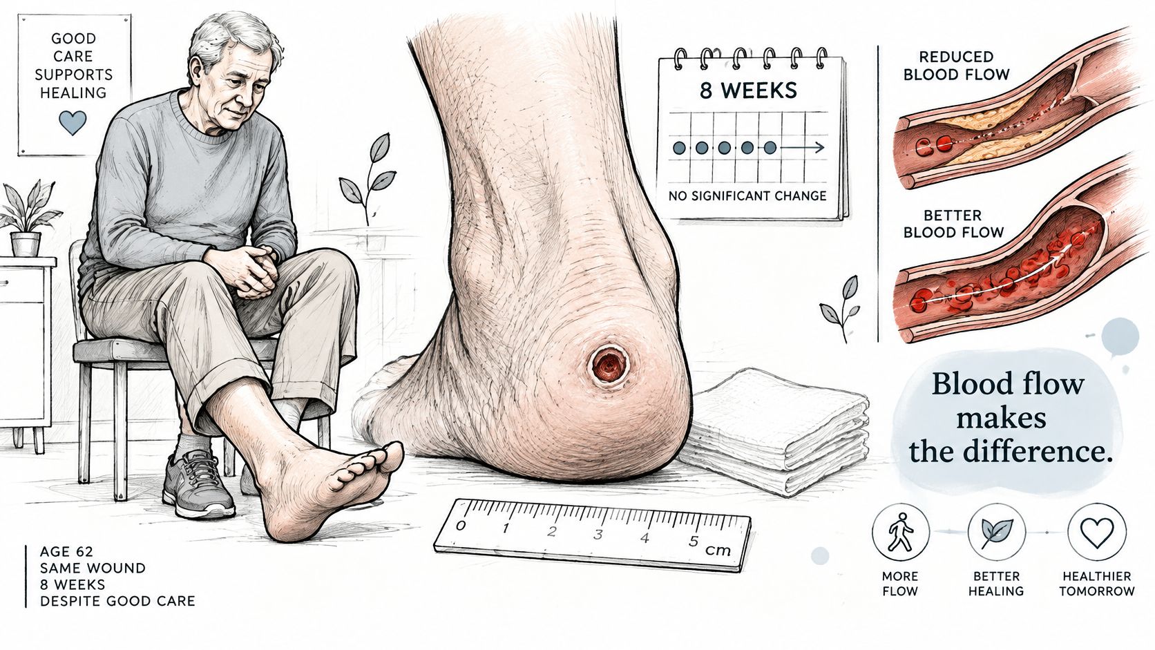
Consider a hypothetical patient, a 62-year-old with a heel ulcer that's been open for eight weeks. The dressing stays in place, there's little drainage, and the wound looks quiet. Yet its measurements haven't changed.
A wound is like a construction site, and blood vessels are its supply routes. Repair requires oxygen, nutrients, and immune cells. Peripheral artery disease (PAD) narrows or blocks arteries, so those supplies may not reach the foot in sufficient amounts, even when the surface looks clean.
The illustration below shows why a quiet-looking heel ulcer can still need circulation testing.
A wound can be the first warning
A review in Circulation Research estimated that 236 million adults worldwide had PAD in 2015, a global prevalence of 5.6%. PAD can cause no obvious symptoms or symptoms that don't resemble classic walking-related leg cramps. The absence of cramping therefore doesn't make a persistent foot wound reassuring.
Chronic limb-threatening ischemia (CLTI) is advanced arterial disease associated with ischemic rest pain, nonhealing ulcers, or gangrene. Ischemia means tissue isn't receiving enough blood. Rest pain means pain from inadequate circulation even when the person isn't walking.
This isn't a diagnosis patients should make from a wound photograph. A clinician needs to connect the wound findings with objective circulation testing and the overall medical picture.
Important distinction: A clean wound surface tells you something about local care. It doesn't prove that the tissue underneath has enough circulation to heal.
Why stalled healing deserves timely attention
The stakes extend beyond wound closure. A review of CLTI outcomes reported one-year mortality ranging from 15% to 40% across studies of patients with CLTI. Those figures describe advanced disease populations, not the prognosis of every person with a foot ulcer.
For wound care, peripheral artery disease changes the central question from “Which dressing should we try?” to “Can this tissue receive what it needs to repair?” The practical questions are which perfusion measurements matter, which tests can answer them, and when specialist assessment shouldn't wait.
Perfusion Numbers That Guide Wound Decisions
A heel ulcer can look dry, pink at the edges, and unchanged, yet still have no realistic path to closure if arterial inflow is poor. Perfusion numbers help separate “needs better local care” from “needs blood-flow assessment now.” They are triage tools: each one estimates a different part of the supply route, so the value matters only beside the wound, the limb, and the reliability of the test.
Perfusion values and what they signal
| Measure | Healing potential signal | Urgent referral signal |
|---|---|---|
| Toe pressure | At least 30 mmHg | Below 30 mmHg |
| Skin perfusion pressure | At least 40 mmHg | No standalone urgent cutoff is used; interpret with the wound findings and other perfusion tests |
| Transcutaneous oxygen (TcPO2) | At least 25 mmHg | Below 30 mmHg indicates severe ischemia |
| Ankle pressure | No standalone healing guarantee | Below 50 mmHg |
| Ankle-brachial index (ABI) | Interpret with waveforms and local perfusion tests | Below 0.4 indicates severe ischemia |
For diabetic foot wounds associated with PAD, the IWGDF PAD guideline identifies toe pressure below 30 mmHg, ankle pressure below 50 mmHg, ABI below 0.4, or TcPO2 below 30 mmHg as severe ischemia warranting urgent vascular consultation; its healing assessments also use toe pressure of at least 30 mmHg, skin perfusion pressure of at least 40 mmHg, and TcPO2 of at least 25 mmHg.
Read the result alongside the wound
Each number answers a narrower question. Toe pressure measures pressure in a small artery supplying a toe. Skin perfusion pressure estimates the pressure needed for blood to return to the skin's small vessels. TcPO2 estimates oxygen availability through the skin. ABI compares ankle pressure with arm pressure, so it describes the limb more broadly and can miss a focal problem near the foot.
A result can support healing potential without proving that healing will occur. Infection, pressure, tissue loss, and the patient's overall condition still matter. Likewise, a low value signals concern, not certainty that amputation will occur.
The TcPO2 entries show why these are not simple pass-or-fail grades. A value can offer some favorable prognostic information while still falling inside the range that warrants urgent specialist assessment. Different thresholds answer different questions: one asks whether repair is plausible, the other asks whether arterial disease is severe enough to change the referral timeline.
Look for agreement, not a single reassuring number
Say an ulcer remains unchanged while one bedside result appears acceptable. The next question is whether the test was reliable, whether it reflects circulation near the wound, and whether other findings disagree. A reading close to a decision threshold may need confirmation rather than being rounded into a reassuring category.
Extensive infection or tissue loss can justify escalation even when pressures are not below a listed cutoff. For skin perfusion pressure, use the healing threshold and the clinical pattern rather than hunting for a separate emergency number.
Practical rule: When perfusion is poor, coordinate vascular assessment before elective aggressive debridement or closure. Urgent infection drainage may need to proceed alongside vascular treatment rather than wait.
How to Test Blood Flow in a Stalled Wound
A pulse check is a starting point, not a complete clearance for healing. The diabetic foot ulcer guidance states that physical examination alone doesn't reliably exclude PAD in someone with diabetes and a foot ulcer. Clinicians should combine examination findings with pressure measurements and Doppler waveforms.
A practical testing routine
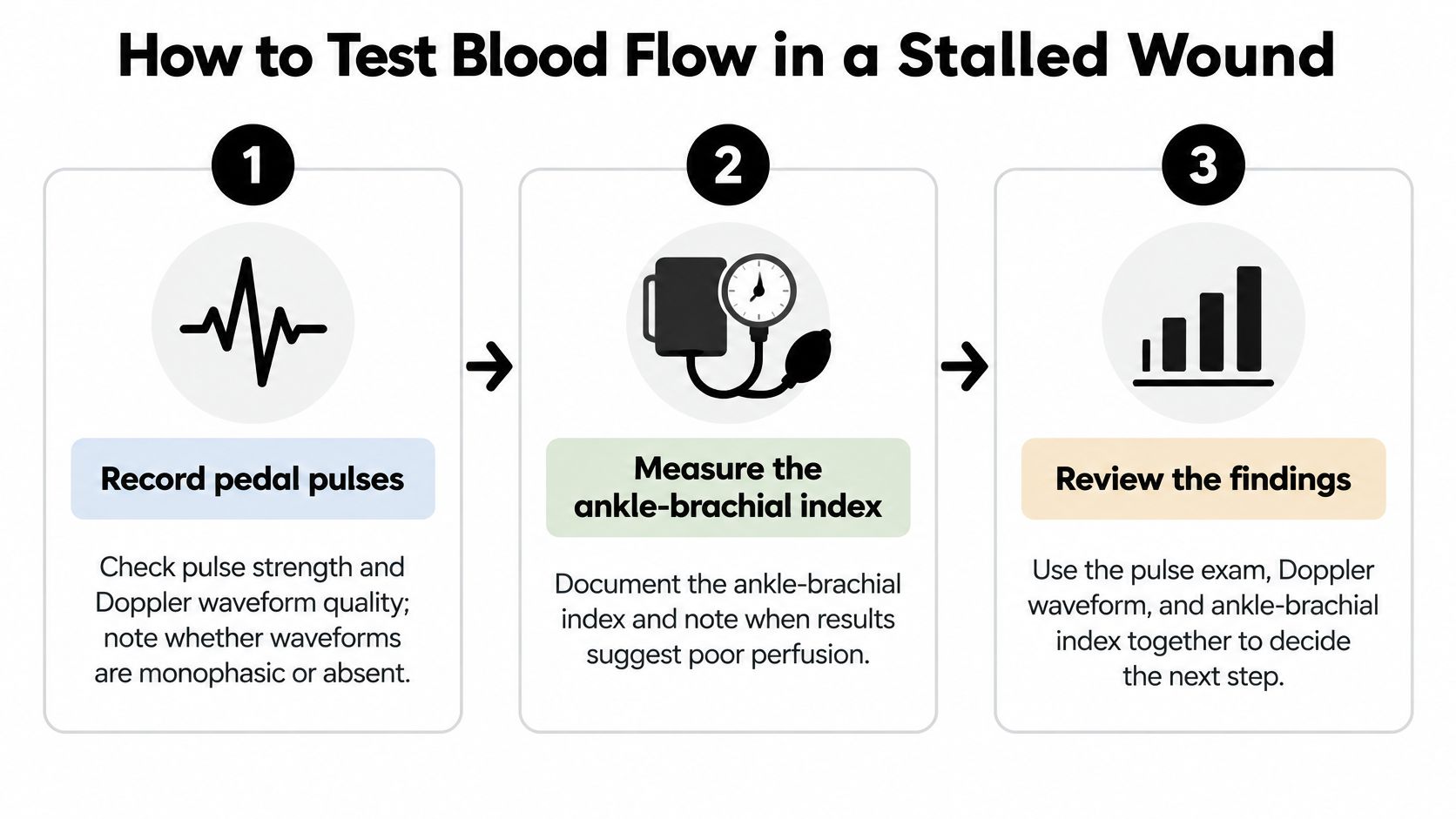
- Record pedal pulses and Doppler waveform quality. The clinician feels for foot pulses and uses a handheld Doppler to assess the arterial signal. A monophasic waveform, meaning a simplified flow pattern, or an absent signal increases concern and deserves explicit documentation.
- Measure the ankle-brachial index. ABI provides a useful comparison between ankle and arm pressures. However, stiff, calcified arteries can resist compression and produce a misleadingly high result, particularly in diabetes, so the report should flag reliability concerns.
- Add toe pressure and the toe-brachial index with waveforms. These tests assess circulation farther down the limb and can help when ankle results are unreliable. Toe measurements are often less affected by arterial calcification, but they still need interpretation rather than being treated as infallible.
- Use TcPO2 or skin perfusion pressure when needed. These tests add information about local oxygen delivery or small-vessel perfusion when ankle and toe findings are unclear or don't fit the wound. The testing location matters because the clinical question concerns the tissue that must heal.
- Arrange vascular review when findings suggest inadequate flow. Pressure tests help establish the physiological problem; arterial imaging helps locate it. The specialist determines the appropriate imaging and whether restoring flow is feasible and likely to help.
- Avoid repeated elective debridement or closure attempts without a perfusion plan. Removing tissue creates an additional healing demand. This doesn't mean withholding urgent treatment for infection, but it does mean coordinating procedures rather than repeatedly treating the surface alone.
The simplified graphic highlights the initial assessment, which may need the additional tests described above.
The common slip is stopping at ABI
An ABI report marked acceptable can seem like the end of the circulation workup. It shouldn't be when the ulcer remains stalled, the arteries may be calcified, or the Doppler findings suggest otherwise. Ask what the result can exclude in this particular foot.
Perfusion tests estimate healing potential; they don't guarantee closure or dictate a procedure by themselves. Clinicians can also use WIfI, which stands for wound, ischemia, and foot infection, to assess the combined threat to the limb and possible benefit of revascularization.
A useful record should include:
- Pulses: Which foot pulses were present or absent.
- Waveforms: The quality of the Doppler signals.
- ABI limitations: Whether calcification or technical factors affected interpretation.
- Toe testing: Toe pressure and associated findings.
- Local perfusion: TcPO2 or skin perfusion pressure when performed.
Offloading, Infection and Diabetes Care Alongside Circulation
Waiting for test results shouldn't mean suspending wound care. The American Heart Association PAD guideline materials support integrating vascular assessment with wound management and restoring flow to the wound bed when clinically feasible. A circulation plan and a local wound plan should work together.
Keep protecting the tissue
Offloading and pressure relief. A heel ulcer requires protection from pressure both in bed and during movement. Depending on the wound, the team may prescribe heel suspension, protective footwear, or a customized offloading device; a dressing alone doesn't remove pressure.
Infection control. Infected tissue needs prompt assessment and treatment. Poor perfusion limits oxygen delivery and immune-cell function and can reduce antibiotic effectiveness, so prescribing medication doesn't remove the need to assess circulation.
Diabetes management. Follow the prescribed glucose-monitoring and medication plan, including any changes needed around procedures or reduced food intake. Don't independently adjust medication because a wound has stalled; the diabetes and wound teams need a coordinated plan.
Avoid treatments that add risk
Compression. Don't start compression for a swollen leg until a clinician has assessed arterial circulation and prescribed an appropriate approach. Mixed arterial and venous disease requires particular care because compression that's unsuitable for the arterial supply can worsen ischemia.
Nutrition and debridement. Maintain adequate protein and fluid intake within any kidney, heart, or other medical restrictions. Leave removal of dead tissue to the treating team, especially when the wound is dry, black, or potentially ischemic.
Our conservative foundation includes dressings, pressure relief, nutrition, and treatment of underlying conditions. The advanced wound care information provides context for how these measures fit within care for complex wounds.
What this looks like at home
Consider a hypothetical patient with diabetes whose heel ulcer has poor toe-pressure findings. The immediate plan isn't to book a procedure and stop everything else. Heel protection, prescribed dressings, infection assessment, and diabetes care continue while urgent vascular review is arranged.
Caregivers should watch for:
- Color or temperature changes: New pallor, blue discoloration, darkening, or a newly cold foot.
- Changing symptoms: Increasing pain or a sudden change in sensation.
- Possible infection: New foul odor, spreading redness, swelling, or increased drainage.
Report these changes the same day. Sudden severe pain, a cold or numb foot, rapidly spreading infection, or systemic illness warrants emergency assessment, not a routine appointment request.
How Coordinated Care Supports Healing After Referral
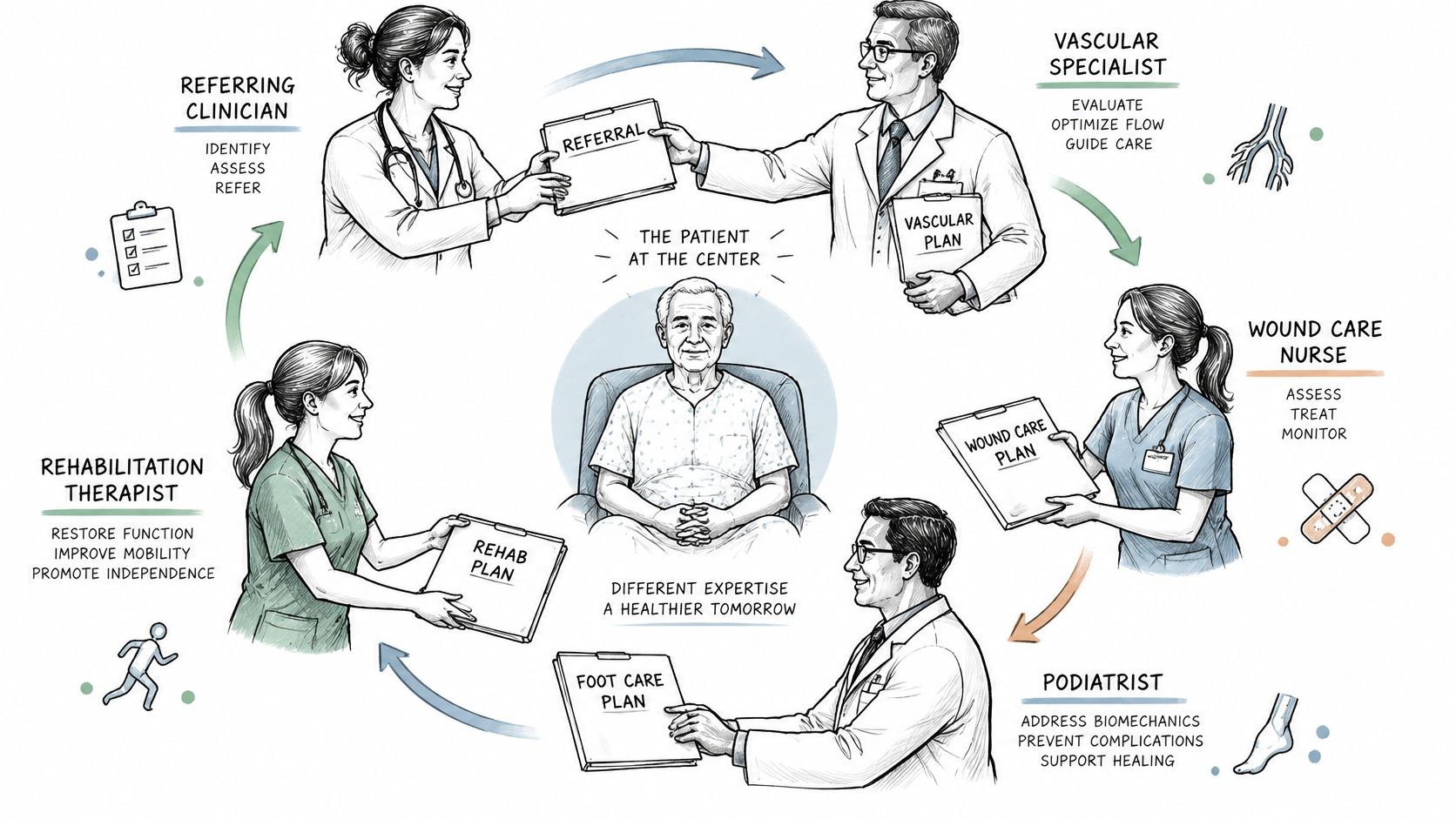
Referral works best as a relay with clear handoffs, not a series of disconnected visits. The patient shouldn't have to carry unanswered questions between offices or discover that nobody has responsibility for the next step.
The patient belongs at the center of that coordination.
Make each handoff explicit
The referring clinician supplies the history and treatment response. A vascular specialist evaluates perfusion and possible revascularization, while a wound-care surgeon assesses debridement or other procedures. A surgical wound-care nurse validates findings, diabetes and infection specialists address those conditions, and home health, skilled nursing, or assisted living staff carry out daily dressings and offloading.
In a 2024 Journal of Vascular Surgery study, early vascular-provider involvement was associated with better wound healing in patients with chronic wounds and PAD, with a hazard ratio of 2.42 (95% confidence interval, 1.21–4.84). That association supports timely involvement, but it isn't a guarantee for an individual patient.
At CWS, we review the history, wound, imaging, and treatment response virtually, then a surgical wound-care nurse validates the wound in person. Our practice addresses fragmented care, long referral waits, and avoidable hospitalizations while keeping existing providers involved.
The accompanying video offers additional context for the care pathway.
Keep one usable plan
Written encounter notes, recommendations returned to existing providers, and follow-up toward closure help prevent conflicting instructions. Our information about coordinated direct care explains the practice's approach.
Before each handoff, caregivers should have:
- Current measurements: Wound dimensions and dated photographs when available.
- Medication list: Including recent antibiotics and medication changes.
- Referral records: Test results, imaging, and specialist recommendations.
- Next review: The appointment date and who to contact if the wound changes.
When Referral Is Needed and What to Ask For
Conservative care isn't passive waiting. It means actively treating pressure, infection, and underlying illness while measuring whether the plan is working. A concerning wound does not need to complete an arbitrary waiting period before specialist evaluation.
The IWGDF guidance on PAD and diabetic foot ulcers says to consider vascular imaging and revascularization when an ulcer with PAD fails to improve within six weeks despite optimal management, regardless of bedside-test results. That is a reassessment trigger. Ischemia, infection, or tissue loss can justify earlier action.
Findings that justify specialist referral
The findings below are part of our escalation pathway for surgical wound evaluation. They prompt assessment, and an evaluation does not automatically lead to surgery.
| Finding | Why It Matters | Next Step |
|---|---|---|
| No measurable size reduction | Healing has stalled despite care | Specialty review of perfusion and treatment response |
| Necrotic or dead tissue | Tissue may lack adequate blood supply | Urgent vascular and surgical assessment |
| Persistent undermining | Tissue loss extends beneath the wound edge | Surgical wound evaluation |
| Infected tissue or recurrent cellulitis | Infection may progress despite local care | Prompt infection control and perfusion assessment |
| Exposed tendon or bone | Deep structures are vulnerable | Surgical evaluation without delay |
| Persistent slough tissue | Nonviable tissue remains in the wound bed | Debridement decision after perfusion assessment |
| Recurrent wound breakdown | The underlying cause may remain unresolved | Review pressure, protection, and vascular status |
| Minimal improvement despite evidence-based care | The current plan needs reassessment | Consider vascular imaging rather than continuing unchanged |
A useful pathway works like a map with decision points: repeat measurements, infection assessment, pressure redistribution, then objective perfusion testing. When indicated, vascular imaging maps the arterial supply from the aorta through the foot arteries, so the team can weigh potential benefit, procedural risk, and the patient's goals.
A bedside number can be normal while the wound still lacks enough usable flow, and a low number cannot by itself choose the procedure. That is why the referral question should name the test, the result date, and the decision the team is trying to make.
Bring a specific request
- Write down the wound history. Include current dimensions, the date measurements began, and treatments already tried.
- Request objective circulation assessment. Ask whether ABI with waveforms, toe pressure, or TcPO2 fits the question and whether existing results are reliable.
- Describe concerning changes immediately. Necrosis, exposed structures, infection, or worsening tissue loss warrants urgent vascular and surgical triage rather than waiting for a routine review.
- Bring the records. Measurements, photographs, medication lists, and test reports help the specialist make an informed decision.
Revascularization improves blood flow and healing potential, but it doesn't replace wound care. Poor perfusion also doesn't mean every patient needs the same procedure.
---
Wound Services offers virtual and in-person evaluation for complex, stalled, and potentially surgical wounds, with care coordinated alongside existing providers. If a foot or leg wound isn't progressing, visit Comprehensive Wound Services to contact our team or refer a patient, and bring the wound measurements, treatment history, and any circulation test results.