
Slough is nonviable, devitalized wound tissue, often cream, yellow, grey or tan, that can cover the wound bed and hide what lies beneath. You're most likely reading about the slough medical term because you've seen it in a wound note, heard it during a dressing change, or spotted a yellowish film and wondered whether it means infection.
That worry is understandable, because the color looks alarming and the word itself sounds like a diagnosis. It isn't. Learning to read slough as a description of tissue, then watching how it changes over time, is far more useful than reacting to a single color on a single day.
## Table of Contents - What Slough Means in Wound Care - What slough looks and feels like - Telling Slough Apart From Eschar, Pus and Granulation - Slough versus pus - Slough versus eschar - Why Slough Does Not Prove Infection - Two wounds, same yellow color - How Clinicians Record Slough at Each Visit - Reading a Slough Entry in a Wound Note - Reading it one field at a time - What the next visit adds - How Slough Can Change a Pressure Injury Stage - Where stage 3 ends and stage 4 begins - A worked example - Why Slough Keeps Coming Back - Look for the driver underneath - When to Get Persistent Slough Reviewed - Bring a record that shows the trend
What Slough Means in Wound Care
A nurse removes a dressing and finds the wound bed covered by a cream colored film. She can't see the base clearly, and the patient asks the question almost everyone asks at this point. Is that infection, dead tissue, or both?
The film she's looking at is likely slough. The Wounds International slough consensus document describes slough as nonviable tissue that can appear cream, yellow, grayish, or tan and may be loose, stringy, slimy, fibrinous, or firmly attached to the wound bed. It's devitalized wound material, meaning tissue and debris that are no longer living or functioning as healthy tissue should.
What slough looks and feels like
Texture often tells you more than color. Some slough lifts away in loose, stringy strands when the wound is gently cleansed. Other slough clings firmly to the bed and doesn't move at all, and the same wound can show both types in different spots.
Think of slough like wet paint that has dried and peeled on a canvas. The peeling layer sits on top and hides the picture underneath, so you can't judge what's below until enough of it is cleared. In the same way, slough can obscure viable tissue, which is the living tissue the clinician needs to see to judge depth and healing.
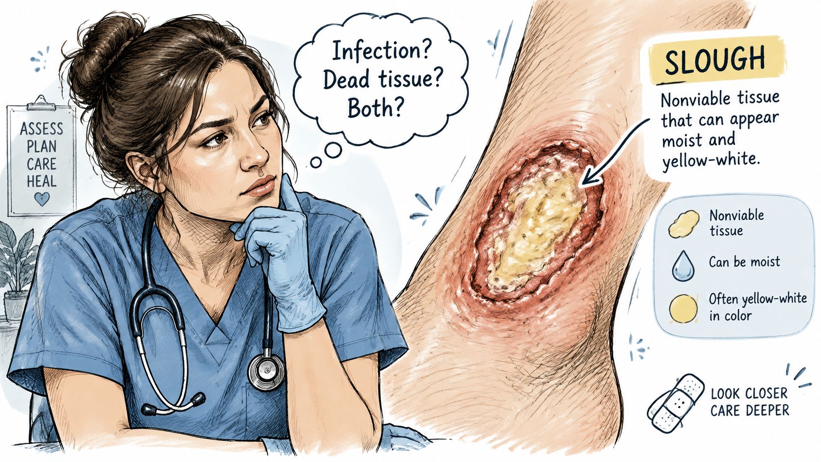
Slough shows up in many kinds of wounds, not just long standing ones. You'll see the term in notes for dehisced surgical wounds, traumatic wounds, skin grafts, diabetic foot ulcers, pressure ulcers, and venous leg ulcers. Finding it doesn't tell you why the wound exists, only what is sitting in it today.
Slough is a finding about tissue, not a diagnosis.
That distinction is the whole point of the term. When you read "slough present" in a note, you now know the writer is describing the wound bed, not naming its cause and not declaring it infected. The cause still has to be worked out from the full picture.
Telling Slough Apart From Eschar, Pus and Granulation
Four findings get confused constantly at the bedside, and the confusion usually starts with color. Yellow doesn't always mean pus, dark doesn't always mean the wound is shallow, and red isn't always a warning sign. The table below puts the four side by side so you can compare appearance, texture, and what each one means.
| Finding | Typical appearance | Texture and attachment | Clinical meaning |
|---|---|---|---|
| Slough | Cream, yellow, grey or tan | Loose, stringy or firmly attached | Nonviable wound material; not an infection marker on its own |
| Eschar | Black, brown or tan, dry | Hard, leathery, firmly adherent | Nonviable tissue that can cover deeper structures |
| Purulence (pus) | Yellow, green or milky fluid | Liquid that drains from the wound | Associated with infection; needs prompt evaluation |
| Granulation tissue | Red or pink, bumpy | Moist, bleeds easily when touched | Viable tissue forming during healing |
Slough versus pus
This is the mix up that causes the most anxiety. Slough is solid material, even when it's soft and stringy, and it sits on or sticks to the wound bed. Purulence is a liquid. It drains, pools in the dressing, and keeps coming because the body is producing it.
Say a yellow film wipes away easily during cleansing and leaves a cleaner bed behind. That behavior points toward loose slough. Say instead that a yellow fluid has soaked through the dressing and collects again within hours. That behavior points toward purulence, and it deserves prompt clinical review because pus is associated with infection.
Slough versus eschar
Eschar is also nonviable tissue, but it behaves differently. It's typically dry, hard, and dark, with a leathery feel, and it's usually stuck down firmly. Slough is generally softer and lighter in color, and it can be moist or stringy.
Both can hide what's underneath, which is why neither should be judged by surface appearance alone. A wound sealed under eschar may look quiet while deeper structures sit just below it. Granulation tissue is the opposite problem. It's red or pink, moist, slightly bumpy, and it bleeds easily when touched, which is exactly what you expect from living, healing tissue with a blood supply.
Practical rule: Match the texture and behavior, not just the color, and let a clinician confirm what you're seeing.
If you're a patient or caregiver, don't try to settle the question by comparing your wound to photos online. Lighting, dressings, and dried exudate all shift colors. Describe what you see, note whether it's solid or liquid, and ask the clinician to name it during the assessment.
Why Slough Does Not Prove Infection
Plenty of simplified advice online says yellow dead tissue must come off right away, as if the color itself were an emergency. That framing skips what slough is. It's a heterogeneous mixture, and its parts vary from wound to wound and week to week.
The Journal of Wound Care debridement consensus notes that slough contains exudate proteins, degraded extracellular matrix material, white blood cells, and microorganisms, so its color, thickness, odor, and attachment vary substantially. Because the mixture varies, its appearance can't show whether the wound is healing. Odor or discoloration on its own is not the same thing as invasive infection.
Two wounds, same yellow color
Consider two wounds that both show yellow slough today. In the first, the wound is a little smaller each week, drainage is lighter, pain is steady, and the skin around the edges looks calm. A clinician reading that trend would likely continue routine care with scheduled review.
In the second, the wound is growing, redness is spreading into the surrounding skin, pain is climbing, and the patient has a fever. That wound needs prompt specialist review. Both wounds have slough, so the slough itself isn't what separates them. The trend and the systemic signs do.
Clinicians judge that trend across visits rather than at one glance. They track wound size, depth, undermining, drainage, pain, the surrounding skin, systemic symptoms, perfusion, pressure and offloading, and how the wound responds to treatment. A single visit gives a snapshot, and snapshots mislead.
Judge the trend, not the colour.
There's a safety reason for slowing down here. People who read that slough must be removed sometimes try aggressive self removal at home, which can damage viable tissue and cause bleeding and pain. Sharp debridement belongs with a competent healthcare professional, and the right method depends on perfusion, pain, bleeding risk, goals of care, and how much slough there is and how firmly it's attached.
How Clinicians Record Slough at Each Visit
Consistent notes are what make trend spotting possible. If one clinician writes "yellow stuff" and the next writes "looks infected," nobody can tell whether the wound changed or only the wording did. Recording the same fields, the same way, at every visit fixes that.
The checklist below shows the items a complete entry covers.
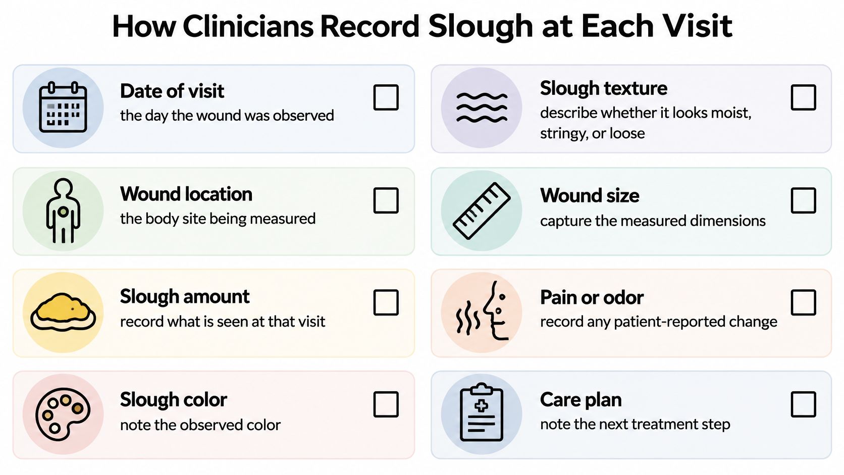
A Journal of Internal Medicine review on wound assessment recommends documenting slough's percentage, depth, consistency, odor, drainage, and location at each assessment. It also describes slough as material that can consist of fibrin, dead cells, and wound exudate, which is why describing what you see matters more than guessing at a cause. An entry should also state whether the slough is new, increased, reduced, or unchanged since the last visit.
The video below walks through how these observations fit into a routine wound assessment.
Once the wound is recorded, the practical steps follow a familiar order. The clinician cleanses the wound to remove surface contaminants and loose debris, protects the periwound skin, then agrees on a debridement approach based on perfusion, pain, bleeding risk, goals of care, and clinician expertise. Cleansing and debridement aren't interchangeable, because cleansing handles the surface while debridement removes devitalized tissue itself.
Between visits, dated photos and measurements make these comparisons far more reliable, which is why many teams use structured tracking such as remote wound monitoring to keep the record consistent. When you read any wound note, run through coverage, size and depth, colour and consistency, attachment, odour, drainage, location, periwound skin, and change since last time. If a field is missing, that gap is worth asking about.
Reading a Slough Entry in a Wound Note
Notes look dense, but a slough entry breaks down into simple pieces once you read it slowly. Say a clinician writes this illustrative line. Slough covers about 40 percent of a 3 cm wound bed, is yellow and moist, is loose, has no odour, and the periwound skin is intact.
Reading it one field at a time
- Coverage at 40 percent: A large part of the base is hidden, so depth underneath that portion can't be confirmed yet.
- Yellow, moist, and loose: Solid material with a soft texture fits slough better than pus, which would drain or pool as liquid.
- No odour: Nothing in this field suggests deterioration at this visit, though odour alone never settles the infection question.
- Periwound skin intact: The surrounding skin shows no spreading redness or breakdown right now.
Notice what the entry doesn't claim. It doesn't say the wound is healing, and it doesn't say treatment has failed. One entry is a single frame, and you need at least two frames to see movement.
What the next visit adds
Now say the next illustrative entry reads differently. Slough covers about 15 percent of the bed, the wound measures 2.5 cm, odour is absent, and the periwound skin remains intact. Coverage has fallen and size has shrunk, so the direction looks favourable. You can see that only because both entries recorded the same fields in the same way.
Reverse the example and the same fields tell the opposite story. If coverage rose instead, the slough became firmly adherent again, odour appeared, and redness spread into the periwound skin, those changes would signal reassessment rather than routine care. Nothing about the word slough changed. The numbers and descriptors around it did.
For each field in the note, ask one question. Did it change since last time, and in which direction?
That habit turns a confusing note into a story you can follow. Coverage going down while size shrinks is progress you can point to. Coverage climbing while pain and drainage increase is a pattern worth raising at the next visit, with the dated entries in hand.
How Slough Can Change a Pressure Injury Stage
Pressure injury staging depends on a deceptively simple requirement. The clinician has to see the deepest tissue involved. When slough covers the base, that requirement can't be met, and guessing a stage from a hidden base helps nobody.
The AHRQ pressure injury chartbook explains that under NPIAP based staging, a wound is classified as unstageable when full thickness skin and tissue loss is present but the wound base cannot be confirmed because slough or eschar obscures it. Unstageable isn't a mild category. It's an honest statement that depth is currently unknowable.
Where stage 3 ends and stage 4 begins
Once enough devitalized tissue is removed to show the base, the injury may turn out to be stage 3 or stage 4. Stage 3 can include visible adipose, which is fat, along with granulation tissue, epibole, undermining, tunneling, and slough. Fascia, muscle, tendon, ligament, cartilage, and bone are not exposed at that stage.
Stage 4 crosses a clearer line. Deep structures such as muscle, tendon, or bone are exposed or directly palpable, meaning the clinician can feel them, and slough may still be present in parts of the bed. The stage follows the deepest tissue seen, not the average appearance of the wound.
A worked example
Say a 3 cm sacral wound is recorded as unstageable because slough covers about 60 percent of the base in this illustrative case. After gentle debridement by the clinician, the base shows adipose tissue and no deeper structures. The wound is then recorded as stage 3.
The injury didn't get worse during that appointment. Removing slough reveals depth, it does not create it. Families sometimes hear the new stage as bad news about care, when it's really new information about anatomy that was there all along.
Good documentation supports that sequence. Clinicians record whether the wound is completely or partly covered, and they avoid assigning a shallower stage from a partly hidden base. Escalation makes sense when slough appears alongside undermining, tunneling, exposed structures, recurrent infection, malodor with deterioration, or stalled healing.
Why Slough Keeps Coming Back
A caregiver cleans a heel ulcer carefully on Monday and feels encouraged. By Wednesday there's a shiny, slimy layer again, and it looks as if Monday's work accomplished nothing. That experience is common, and it doesn't automatically mean the cleaning was done badly.
Debridement is rarely a one time step, because a wound surface can reform quickly when ongoing intervention is absent. A 2025 review in PMC describes a shiny or slimy layer that may recur within 1 to 2 days, while also noting limited consensus around which clinical signs reliably identify biofilm. Biofilm is a community of microorganisms embedded in a protective matrix, and slough can act as a physical scaffold that supports it.
That scaffold matters for treatment. The surrounding matrix may protect organisms from immune clearance and reduce the effectiveness of topical treatment, so the layer keeps rebuilding itself. Still, recurrence is not proof of a specific organism, and it isn't proof of systemic infection either.
Look for the driver underneath
When slough returns quickly, clinicians check for unresolved drivers rather than merely repeating removal. Common ones include:
- Pressure or poor offloading: Ongoing load keeps damaging tissue faster than it can recover.
- Ischemia and edema: Limited blood flow and excess fluid both slow the clearing of debris.
- Excess moisture or dead space: Fluid collects where it shouldn't, and material accumulates with it.
- Foreign material or deep infection: Something below the surface keeps feeding the problem.
- Nutrition and diabetes control: Inadequate nutrition and uncontrolled diabetes undermine repair throughout the body.
The honest caveat is that evidence here is still developing, and high quality randomized trials are scarce for some wound cleansing and biofilm practices. That's why plans are individualized instead of copied from a single protocol, and why advanced wound care focuses on reassessing the whole wound and its drivers. Repeatedly traumatizing tissue at home can cause pain and damage viable tissue without fixing any driver on that list.
When to Get Persistent Slough Reviewed
Some patterns justify asking for a specialist look rather than another round of the same care. We list eight indicators that a wound may deserve surgical evaluation, and several involve slough directly or the company it keeps.
- No measurable size reduction: The wound holds steady or grows despite appropriate care.
- Necrotic or dead tissue: Nonviable material persists in the bed.
- Persistent undermining: Tissue separates under the edges and doesn't close down.
- Infected tissue or recurrent cellulitis: Infection keeps returning after treatment.
- Exposed tendon or bone: Deep structures are visible or palpable.
- Persistent slough tissue: Slough reaccumulates shortly after treatment, again and again.
- Recurrent wound breakdown: The wound closes, then reopens.
- Minimal improvement despite appropriate evidence based care: Good care isn't producing change.
Separate those review triggers from urgent warning signs. Increasing pain, spreading redness, fever, or worsening drainage need same day medical contact, not a wait for the next routine appointment. Those signs suggest the situation is moving, and moving problems shouldn't sit in a queue.
Bring a record that shows the trend
A review goes faster when you arrive with dated evidence. Start a baseline entry with coverage, length, width, and depth, then photograph the wound with consent where local policy allows, using the same angle and lighting each time. Note the date of each cleansing or debridement and how the wound looked over the following days.
Our Surgical Wound Evaluation service exists for exactly this kind of reassessment, and our escalation pathway explains how a stalled wound moves from routine care to specialty review. A review doesn't by itself mean surgery. The clinician recommends the next step based on your record and the examination, and your careful notes are what make that recommendation accurate.
---
If slough keeps returning, your wound has stopped shrinking, or you're unsure whether what you're seeing needs prompt attention, we can review your dated measurements, photos, and treatment history through a virtual or in person evaluation and coordinate next steps with your existing care team. Visit Comprehensive Wound Services to ask about a Surgical Wound Evaluation and how reviews are arranged, and bring your wound record so we can judge the trend together.