
If a wound keeps reopening despite careful dressing changes, is the dressing really the problem? Often, the more important question is whether pressure, poor circulation, infection, or an incorrect diagnosis is preventing repair.
The main types of non healing wounds need different treatment plans because they develop for different reasons. For patients and caregivers, understanding those differences helps turn a frustrating cycle of dressing changes into a clearer plan for assessment, treatment, and reassessment.
## Table of Contents - Types of Non Healing Wounds Compared at a Glance - Why appearance isn't enough - Why Each Wound Type Stops Healing - Different causes need different solutions - Infection can complicate the original mechanism - How Wound Types Are Graded and Classified - Pressure injury labels describe tissue damage - Diabetic foot tools answer different questions - Diabetic Foot Ulcers Carry the Heaviest Long-Term Burden - Closure doesn't remove the original risk - Coordinate the whole plan - Venous Leg Ulcers and Pressure Injuries Across Care Settings - What the venous ulcer row means at home - What the pressure injury row means in a facility - How Care Changes by Wound Type - Protect first, then support repair - Checkpoints That Show a Wound Is Not Healing and Next Steps - Interpret measurements alongside the diagnosis - Act on a stalled or unusual wound
Types of Non Healing Wounds Compared at a Glance
A nonhealing wound is one that stalls or repeatedly reopens despite appropriate care. Clinicians assess its progress by its healing trajectory and underlying cause, not just by counting how many days it's been open.
The common categories are diabetic foot ulcers, venous leg ulcers, arterial ulcers, and pressure injuries. Atypical wounds form another group, including ulcers caused by inflammatory disease, unusual infections, or malignancy.
Main types of non healing wounds
| Wound type | Who it mostly affects | Main cause | Typical location and look |
|---|---|---|---|
| Diabetic foot ulcer | People with diabetes, especially with neuropathy | Neuropathy, repetitive pressure, infection, and poor blood flow | Sole, heel, or toe; often painless |
| Venous leg ulcer | Adults with chronic venous disease or past deep vein clots | Chronic venous hypertension | Shin and ankle; shallow, with swelling and skin changes |
| Arterial ulcer | People with peripheral artery disease | Inadequate arterial blood flow | Toes, heels, or foot edges; may be pale, deep, and painful |
| Pressure injury | People with limited mobility in hospitals, nursing homes, or at home | Sustained pressure and shear over bone | Sacrum, heels, or hips; can start as discolored intact skin |
| Atypical wound | Anyone, less commonly | Inflammatory, infectious, vasculitic, metabolic, or malignant disease | Doesn't match the common patterns |
Read across the table rather than choosing a diagnosis from the final column. The person's medical history, likely mechanism, location, and appearance need to fit together.
Why appearance isn't enough
A shin ulcer may involve venous disease, arterial disease, or both. A heel wound could result from loss of sensation, pressure from lying in bed, inadequate circulation, or overlapping problems.
The cause column matters more than the appearance column. Pain isn't a reliable sorting tool either, because someone with nerve damage may not feel a serious wound.
Practical rule: Use the table to organize questions for an assessment, not to choose compression, remove dead tissue, or diagnose a wound at home.
If several categories seem to fit, that's useful information rather than a failed attempt to identify the wound. Mixed causes require a plan that addresses each contributor.
Why Each Wound Type Stops Healing
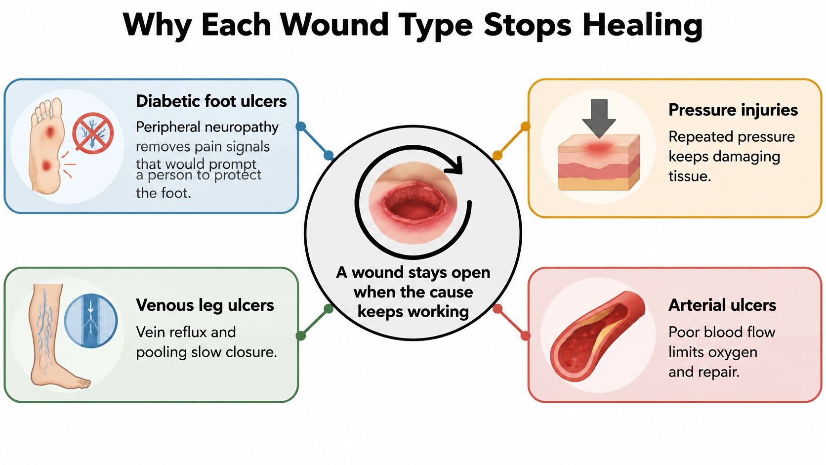
Think of each wound as a fire supplied by a different fuel line. Treating the surface without stopping the ongoing damage is like extinguishing visible flames while fuel continues to flow.
A dressing can protect tissue and manage drainage. It can't, by itself, restore arterial circulation or remove the pressure of walking on an injured foot.
The diagram separates these ongoing causes.
Different causes need different solutions
With a diabetic foot ulcer, peripheral neuropathy reduces protective sensation. A person may continue walking on a rubbed toe or overloaded area of the sole because it doesn't hurt. Repeated loading injures the tissue further, while infection and peripheral artery disease may make repair harder.
A venous leg ulcer develops when blood doesn't return efficiently through the leg veins. Persistent venous pressure contributes to swelling and skin damage. Even excellent dressing care may produce limited progress if the leg remains swollen and the underlying venous problem isn't addressed.
An arterial ulcer involves insufficient blood delivery. Repairing tissue needs oxygen and nutrients, and an underperfused foot may not receive enough of either. This is different from venous pooling, even though both conditions involve circulation.
With a pressure injury, sustained pressure and shear damage tissue, often over a bony prominence. Someone sliding down in bed can experience shear as deeper tissues move differently from the skin. Moisture, poor nutrition, impaired sensation, and difficulty repositioning can add to the problem.
Infection can complicate the original mechanism
Infection may coexist with any of these wounds, but it shouldn't become the default explanation for every stalled ulcer. Clinicians assess the wound, surrounding skin, symptoms, and treatment response before deciding whether sampling or additional testing is useful.
Questions about molecular testing belong within that clinical assessment, rather than replacing an examination of pressure and blood flow.
- Repeated injury: Check what touches or loads the wound during walking, sitting, transfers, and sleep.
- Circulatory barriers: Distinguish venous congestion from inadequate arterial supply.
- Unexplained deterioration: Reconsider the diagnosis instead of automatically adding another topical product.
The mechanism determines the treatment sequence. Two wounds that look similar may need very different care.
How Wound Types Are Graded and Classified
A wound's name describes its category, but classification helps describe its seriousness. A useful assessment records tissue damage, infection, and circulation separately, because a small opening can conceal a significant problem.
Patients sometimes assume that every ulcer receives a stage. In practice, pressure injuries use staging, while diabetic foot ulcers often require several complementary classification tools.
Pressure injury labels describe tissue damage
Pressure injuries range from stage 1 through stage 4, progressing from intact skin changes to increasingly deep tissue loss. Clinicians also use unstageable when slough or eschar hides the wound base, preventing them from determining its full depth.
A deep-tissue pressure injury may appear as persistent deep discoloration, including purple or maroon tissue, even when the skin hasn't opened. Intact skin therefore doesn't necessarily mean that the tissue underneath is healthy.
- Visible depth matters: The clinician documents which structures are involved.
- A covered base creates uncertainty: Dead tissue can obscure the extent of injury.
- Discoloration needs assessment: Caregivers shouldn't wait for an opening before reporting a concerning area over bone.
Diabetic foot tools answer different questions
SINBAD records site, ischemia, neuropathy, bacterial infection, area, and depth. It gives clinicians a structured way to describe the ulcer.
The IDSA/IWGDF infection classification addresses infection severity. WIfI, meaning wound, ischemia, and foot infection, helps assess limb threat and the likely benefit of restoring blood flow.
These tools aren't interchangeable. A wound description doesn't establish how much blood reaches the foot, and an infection label doesn't describe all the tissue loss.
Venous and arterial ulcers also require documentation of dimensions, depth, surrounding skin, and vascular findings. Calling an ulcer chronic without identifying its circulatory mechanism leaves an important part of the assessment unfinished.
For an atypical wound, classification begins with the suspected cause: inflammatory, vasculopathic, infectious, metabolic, genetic, or malignant. Testing may then narrow that category to a specific diagnosis.
Exposed tendon or bone, recurrent infection, and suspected ischemia require escalation beyond routine dressing care. A classification is useful only when the care team acts on what it reveals.
Diabetic Foot Ulcers Carry the Heaviest Long-Term Burden
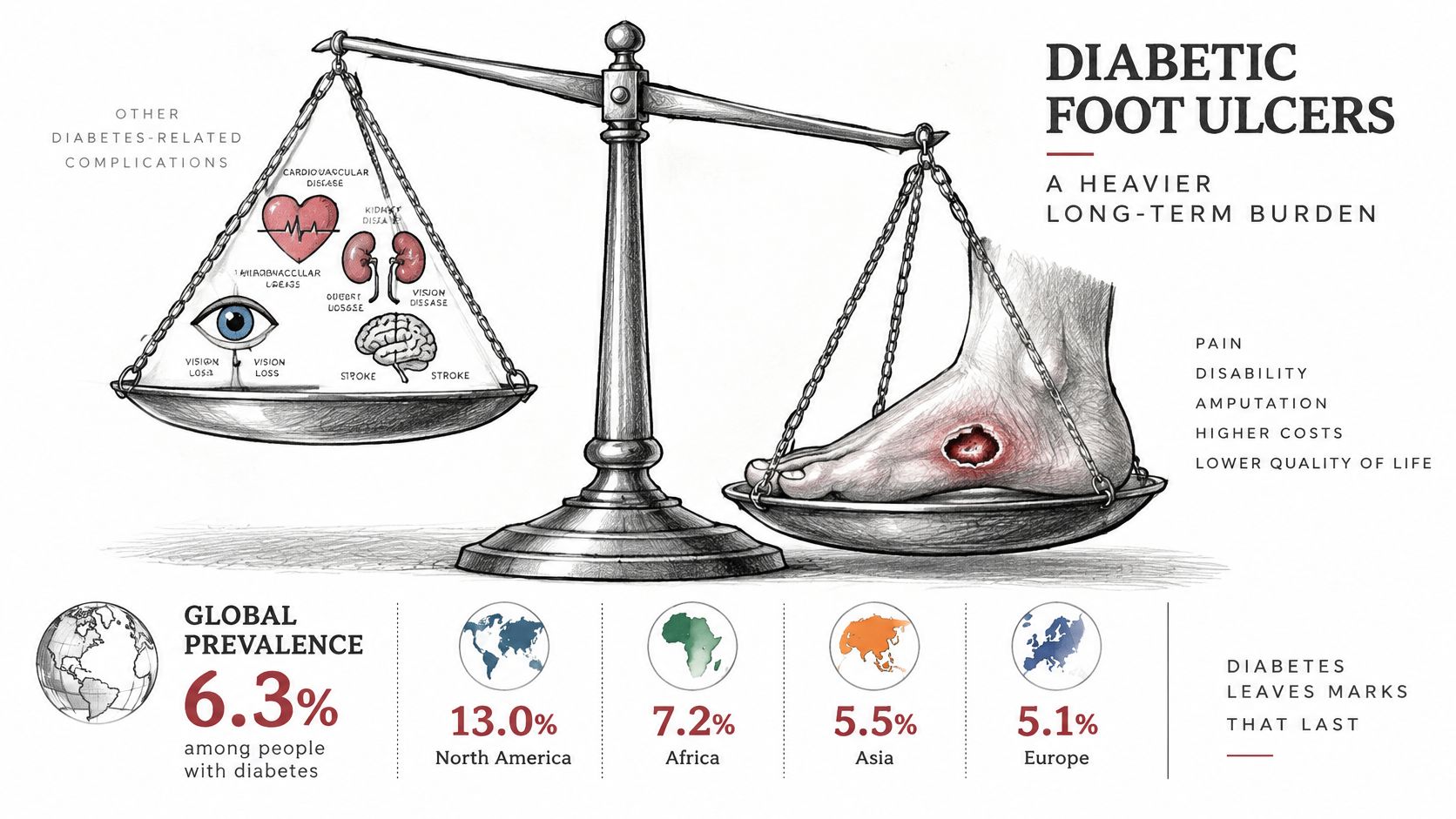
Diabetic foot ulcers deserve attention beyond the visible opening. A systematic review and meta-analysis of diabetic foot ulcer prevalence estimated global prevalence at 6.3% among people with diabetes, with 13.0% in North America.
Those estimates describe populations, not an individual person's prognosis. Their practical importance is that diabetic foot wounds are a substantial complication requiring organized prevention, treatment, and follow-up.
The infographic highlights their long-term significance.
Closure doesn't remove the original risk
Diabetes-related vascular disease, loss of protective sensation, infection, and impaired tissue repair can occur together. Closing the opening doesn't necessarily correct the foot mechanics or restore sensation.
Say a heel ulcer closes after treatment. Later, a new shoe rubs the same spot, but the person doesn't feel the irritation and continues wearing it. The skin can break down again even though the earlier wound received appropriate care.
This is why follow-up includes more than checking whether a dressing is still needed.
- Footwear assessment: Look for rubbing, seams, tight areas, or pressure points.
- Skin inspection: Check the sole, heel, toes, and spaces between toes, using a mirror or caregiver assistance when needed.
- Ongoing protection: Continue the prescribed pressure-relief and footwear plan rather than returning immediately to whatever was worn before.
- Early reporting: Tell the care team about a blister, new drainage, discoloration, or recurrent opening.
A closed wound is a milestone. For a foot with persistent neuropathy or vascular disease, prevention remains part of treatment.
The accompanying video provides another way to engage with wound-care education.
Coordinate the whole plan
A stalled diabetic foot wound needs a coordinated review of perfusion, pressure relief, infection, and metabolic control. Improving blood glucose management is important, but it doesn't substitute for protecting the ulcer from repeated loading or addressing inadequate circulation.
Caregivers can help by keeping the off-loading plan practical. If a prescribed device makes transfers difficult or the patient can't use it safely, report that problem rather than abandoning it without telling anyone.
Surgical options may be appropriate when tissue damage, infection, or other findings warrant intervention. An evaluation should establish what needs correcting and how that correction fits into the ongoing care plan, not assume that every diabetic ulcer requires an operation.
Venous Leg Ulcers and Pressure Injuries Across Care Settings
Venous ulcers and pressure injuries affect different populations, so families and care teams need to plan for different daily challenges. A person who walks independently but has chronic leg swelling may need sustained venous management, while someone unable to turn in bed needs reliable pressure redistribution.
The British Journal of Dermatology wound-burden review describes the population burden and setting-specific estimates summarized below.
Burden of venous leg ulcers and pressure injuries
| Wound type | Prevalence or incidence | Population or cost burden | Range by care setting |
|---|---|---|---|
| Venous leg ulcer | Pooled prevalence 0.32%; pooled incidence 0.17% | 500,000–600,000 people in the US; nearly $1 billion annually; UK £400–600 million | Not reported as a setting range |
| Pressure injury | Setting-specific prevalence estimates | Recurring burden across hospitals, long-term care, and home care | Acute-care hospitals 1%–27%; aged care 7%–53%; community 6%–29% |
These pressure-injury ranges aren't directly interchangeable. Different definitions, patient populations, and surveillance methods mean that the table shouldn't be used to rank an individual hospital against a nursing home.
What the venous ulcer row means at home
Venous ulcer care has to fit everyday life. Compression, when appropriate after arterial assessment, works alongside edema control, mobility, and management of the underlying venous disease.
If swelling persists despite the prescribed plan, the response shouldn't be a more absorbent dressing alone. The team needs to review whether compression fits correctly, whether the patient can use it consistently, and whether another circulatory problem is contributing.
Practical obstacles matter. Someone who can't reach their ankle may need help applying the prescribed system rather than another set of written instructions.
What the pressure injury row means in a facility
Hospitals and residential care settings should assess skin and pressure-injury risk at admission and revisit the plan when mobility or health changes. Existing redness or discoloration should be documented so the receiving team knows what requires attention.
- During transfers: Communicate wound location, support-surface needs, and positioning restrictions.
- During routine care: Check areas that the patient can't see or feel.
- After discharge: Make sure the home caregiver understands how to protect affected areas and whom to contact about changes.
Setting predicts practical needs, not certainty about wound type. A mobile person can develop a pressure injury, and a nursing-home resident can have a venous ulcer. The individual assessment still determines care.
How Care Changes by Wound Type
The most harmful mistake can be applying a useful treatment to the wrong diagnosis. Compression helps many venous ulcers, but applying it without assessing arterial supply can worsen an ischemic wound.
Treatment sequence matters. Establish circulation, identify infection and tissue damage, and remove the ongoing mechanical cause before relying on a more advanced dressing.
Core care, common mistake and escalation trigger by wound type
| Wound type | Core treatment | Mistake to avoid | Escalate when |
|---|---|---|---|
| Diabetic foot ulcer | Off-loading, infection control, glycemic management, and perfusion assessment | Walking on an unprotected wound or assuming neuropathy is the only cause | Exposed structures, recurrent infection, ischemia, or inadequate measured progress |
| Venous leg ulcer | Compression after confirming adequate arterial supply, edema control, elevation, and mobility | Starting compression without vascular assessment | Swelling or stalled healing persists despite the prescribed plan |
| Arterial ulcer | Vascular assessment, consideration of revascularization, and coordinated wound care | Treating inadequate circulation with dressings alone | Rapid deterioration, rest pain, or gangrene |
| Pressure injury | Pressure redistribution, nutritional assessment, moisture control, and debridement when indicated | Leaving pressure on the injured area | Deep tissue damage, an obscured base, or infection is suspected |
| Atypical wound | Diagnostic assessment and wound-edge biopsy when indicated | Repeating empiric treatments without establishing a cause | The appearance or response doesn't fit the working diagnosis |
Protect first, then support repair
For pressure injuries, remove or redistribute the causative pressure while addressing moisture, nutrition, and wound-bed needs. Debridement, which removes nonviable tissue, requires clinical judgment about circulation and the underlying diagnosis.
The IWGDF guideline compilation describes negative-pressure wound therapy as an adjunct for stage 3 and 4 pressure injuries and identifies more than 50% wound-area reduction by four weeks in diabetic or mixed wounds as associated with healing at 12 weeks.
Neither a device nor a benchmark replaces individualized assessment. Negative-pressure therapy doesn't eliminate the need for pressure redistribution, and slower-than-expected progress should prompt reassessment rather than blame.
Treatment safeguard: Don't start stronger compression or remove black tissue yourself because an ulcer resembles a photograph online.
Advanced wound care should address an identified barrier to healing, not add complexity to a plan whose diagnosis remains uncertain.
Before a specialist visit, prepare a written treatment history. Include the dressings, compression systems, off-loading devices, antibiotics, procedures, and test results already tried, along with dates and observed responses.
Photographs and measurements are useful when they're clearly dated. Tell the team what wasn't practical, too, such as a device that couldn't be worn safely or a compression system that repeatedly slipped.
Checkpoints That Show a Wound Is Not Healing and Next Steps
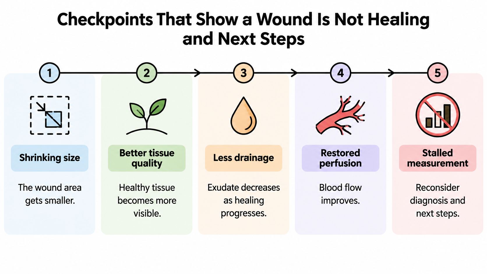
Closure isn't the only evidence of healing. Shrinking dimensions, healthier tissue, less drainage, and improved perfusion can indicate progress, while an unchanged measurement calls for closer review.
These checkpoints help patients and caregivers describe changes clearly.
Interpret measurements alongside the diagnosis
The lower-extremity arterial-disease wound guideline recommends evaluating infection or biofilm and screening for arterial disease when a wound hasn't improved after 4–6 weeks of appropriate care. Deterioration warrants review sooner, not a wait for that interval to end.
Checkpoints and benchmarks for a stalled wound
| Checkpoint | Benchmark | What it signals |
|---|---|---|
| Wound area reduction | Meaningful shrinkage on serial measurements in diabetic or mixed wounds | A flat trajectory requires review of diagnosis, off-loading, and other barriers |
| Arterial reassessment | Worsening or absent improvement during appropriate care | Consider ankle-brachial index, Doppler waveforms, and toe pressure |
| Infection or biofilm check | Persistent lack of progress despite appropriate care | Evaluate infection and screen for arterial disease |
| Skin-perfusion pressure | At least 40 mmHg | Associated with improved healing potential |
| Toe pressure | At least 30 mmHg | Associated with improved healing potential |
| Transcutaneous oxygen pressure | At least 25 mmHg | Associated with improved healing potential |
The perfusion thresholds in the table come from the IWGDF peripheral artery disease guideline; they indicate improved healing potential, not a guarantee of closure.
For a hypothetical measurement example, compare a wound that becomes substantially smaller with one whose dimensions barely change. Both remain open, but they shouldn't automatically receive the same next step.
Act on a stalled or unusual wound
- Record a baseline. Note the wound location, suspected type, start date, and clinician-measured dimensions.
- Track consistently. Use dated measurements and photographs taken under similar conditions; don't probe the wound yourself.
- Report circulation concerns. A cold, pale foot, pain at rest, or rapid deterioration needs prompt assessment.
- Question a poor fit. An unusual wound may need a biopsy rather than another topical treatment.
The American Family Physician chronic-wound assessment guide supports biopsy for atypical nonhealing wounds, including sampling the wound edge to help identify the underlying disease. A wound specialist can arrange this while coordinating ongoing protective care.
Our escalation pathway considers absent size reduction, dead tissue, undermining, infection or recurrent cellulitis, exposed tendon or bone, persistent slough, recurrent breakdown, and minimal improvement despite appropriate care. We review history, the wound, imaging, and treatment response virtually, followed by in-person validation by a surgical wound-care nurse.
Conservative treatment remains the foundation. When intervention is appropriate, the surgeon who evaluates the wound also operates and follows healing afterward.
---
Wound Services offers virtual and in-person specialty evaluation for complex, stalled, and recurrent wounds, with surgical treatment considered only when clinically appropriate. If a wound isn't progressing or keeps reopening, visit Comprehensive Wound Services to request an evaluation and bring your treatment history, recent photographs, and available measurements.