The most popular advice about skin substitutes for wound healing sounds simple. If a wound isn't closing, add an advanced product and it will heal faster. That advice skips the harder questions. What's causing the wound, does it have enough blood flow, is infection controlled, and has pressure been relieved? A skin substitute can't answer those questions for you. It's one tool inside a wound plan, and it only works when the plan around it is sound.
## Table of Contents - What Skin Substitutes Are and Are Not - Why the name causes confusion - Three Product Categories and How Each Works - What each category does - The reality check - Which Wounds Respond to Skin Substitutes - What the evidence does not show - How to Fit a Skin Substitute Into the Wound Plan - A step by step sequence - When a Wound Needs Surgical Evaluation - Why a covering alone cannot fix some findings - Documentation That Decides Coverage and Fit - How coverage rules differ from clinical fit - Confirm Your Next Step With a Wound Specialist - What a specialist review looks like
What Skin Substitutes Are and Are Not
Many patients hear skin substitute and picture new skin placed over a wound, ready to take root. That's the misunderstanding behind most questions. Most of these products aren't transplanted living skin moved from one part of the body to another. They provide a scaffold or wound covering that your body uses to rebuild tissue, which means they don't automatically replace surgical grafting.
A useful analogy is a trellis. A trellis isn't a new plant. It gives a plant structure to grow across, and the plant still needs water, light, and healthy roots. A skin substitute works in a similar way. It supports growth, but blood flow, infection control, and pressure relief still decide whether that growth can happen.
Why the name causes confusion
Regulation reflects this variety. As a Fallon Health policy on skin substitutes notes, FDA does not formally use "skin substitute" as a product-class name. Products may instead be regulated as human cells, tissues, and cellular and tissue-based products, 510(k) devices, or premarket-approved devices. They differ by source, composition, regulatory pathway, and intended use.
That also means advanced doesn't mean superior for every wound. Evidence is more supportive for selected diabetic foot and venous leg ulcers, while randomized trials have not shown a clinically significant benefit for pressure ulcers over standard care. Clearance or approval doesn't prove that every use is clinically effective or covered by insurance.
Sort options by wound cause, depth, vascular status, infection, and treatment goals, not by brand name. That's how you answer the practical question, why was this recommended for this wound but not another?
A skin substitute is a wound covering with a job to do, not a shortcut past the basics.
Three Product Categories and How Each Works
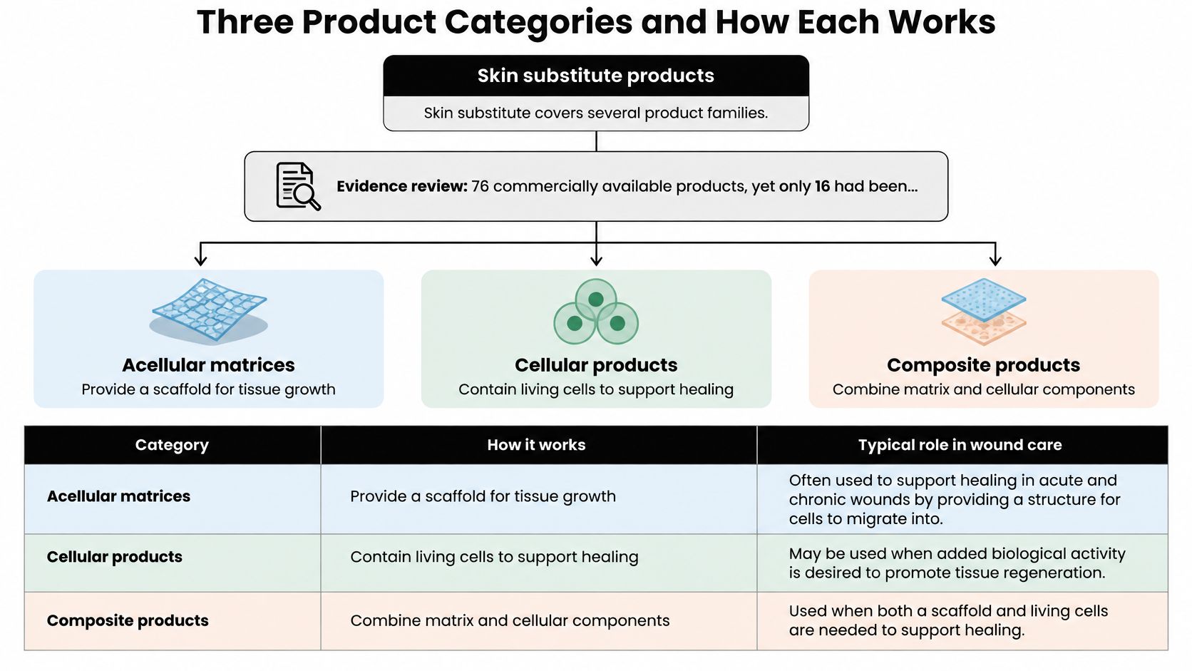
Skin substitute isn't a single technology. It's a label covering several product families that work in different ways, and most haven't been tested in rigorous trials. According to an evidence review of skin substitutes for chronic wounds, researchers identified 76 commercially available products, while only 16 products had been evaluated in randomized trials across diabetic foot, venous leg, or pressure ulcers. That gap is worth remembering when a specific product is proposed.
What each category does
Acellular dermal substitutes contain no living cells. They provide a three-dimensional scaffold for host-cell migration, vascular ingrowth, and extracellular-matrix deposition. Your own cells have to move in and do the building, so perfusion and a clean wound bed matter a great deal.
Cellular dermal substitutes add living cells to a scaffold. Those cells may also deliver paracrine signals that influence inflammation, angiogenesis, and re-epithelialization. Angiogenesis means growth of new blood vessels, and re-epithelialization means new surface skin forming across the wound.
Combined cellular epidermal-dermal constructs join epidermal and dermal layers in one product. The epidermis is the outer skin layer, and the dermis is the deeper supportive layer. Combining them aims to support both coverage and tissue rebuilding.
| Category | How It Works | Biological Activity | Evidence to Ask For |
|---|---|---|---|
| Acellular dermal substitute | Scaffold for host-cell migration, vascular ingrowth and matrix deposition | No living cells; relies on the wound's own cells | Product-specific randomized trial data |
| Cellular dermal substitute | Scaffold that also contains living cells | Living cells may deliver signals that influence inflammation and angiogenesis | Trials in the same wound type and product |
| Combined cellular epidermal-dermal construct | Epidermal and dermal layers combined in one product | Living cells in both layers may support surface closure and dermal rebuilding | Which wound types and comparators were studied |
The reality check
None of the three works on an unprepared wound. Debridement, infection control, moisture balance, pressure redistribution, vascular assessment, and compression for venous disease decide whether a product can function. Debridement is removal of nonviable tissue, and it isn't optional housekeeping. A scaffold placed over dead tissue or uncontrolled infection has little chance to help.
Match the product to etiology, perfusion, depth, exudate, and exposed structures. Etiology means the underlying cause, and exudate is wound drainage. Ask for the trial evidence behind the specific product being suggested. Don't accept the generic label skin substitute as evidence by itself.
Which Wounds Respond to Skin Substitutes
The strongest trial evidence sits with two wound types. A U.S. Agency for Healthcare Research and Quality evidence review identified 18 randomized controlled trials, with 12 involving diabetic foot ulcers and 6 involving vascular leg ulcers. Most trials measured complete wound healing at or before 12 weeks, time to healing, and wound infection. Across the reviewed studies, the majority reported significantly more healed wounds among patients receiving a skin substitute than among patients receiving standard care alone.
That is a real benefit signal for selected wounds. It isn't a promise for every wound, and it doesn't apply equally to every product in the category.
What the evidence does not show
The limits are just as important. Only 7 studies reported wound recurrence, and follow-up beyond the initial healing period was limited. Closure at 12 weeks therefore doesn't establish durable healing. A wound can close and reopen if pressure, venous disease, infection, or diabetes control isn't managed.
Recurrence, infection, off-loading, vascular status, and diabetes control still shape outcomes after the product is placed. Off-loading means taking pressure off the foot, often with a boot, cast, or custom footwear. If a patient keeps walking on a plantar foot ulcer without protection, a substitute can't overcome repeated mechanical stress.
For pressure ulcers, randomized trials have not shown a clinically significant benefit over standard care. Substitutes shouldn't be presented as a first choice there. Pressure relief, repositioning, nutrition, and surface support remain central.
For other complex or stalled wounds, this review does not establish a comparable trial base. That doesn't mean a specialist will never consider a product. It means you shouldn't extrapolate results from diabetic foot or venous leg ulcers to a different wound without a careful assessment.
| Wound Type | Trial Evidence | Role in the Plan |
|---|---|---|
| Diabetic foot ulcers | 12 of 18 trials; majority reported more healed wounds than standard care | Adjunct after offloading and infection control |
| Venous leg ulcers | 6 of 18 trials; majority reported more healed wounds than standard care | Adjunct alongside compression |
| Pressure ulcers | Randomized trials show no clinically significant benefit over standard care | Standard care first; substitutes not established |
| Other complex or stalled wounds | No comparable trial base in this review | Specialist assessment before any product choice |
The table can guide expectations, but it can't tell you how an individual product will perform or whether healing will last. Long-term recurrence remains the open question behind most short-term closure results.
How to Fit a Skin Substitute Into the Wound Plan
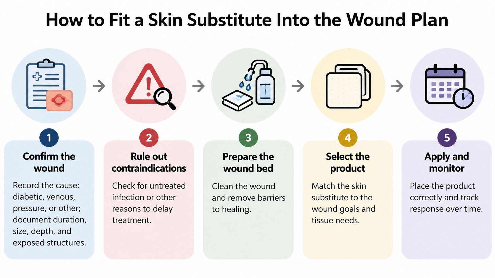
Fitting skin substitutes for wound healing into a plan works best as a sequence, not a single decision. Comparative rankings can inform that sequence without dictating it. A network meta-analysis of 38 randomized trials involving 3,862 patients ranked placenta-derived tissue products highest for wound-healing probability, with a p-score of 0.90, followed by living-cell skin substitutes at 0.70 and acellular substitutes at 0.56 relative to standard care. Those rankings reflect relative performance across different studies, not proof that one product is universally superior for your wound.
A step by step sequence
- Confirm the wound by recording cause, duration, size, depth, and exposed structures, because product choice depends on what you're treating.
- Rule out contraindications by checking for untreated infection or osteomyelitis and for unassessed ischemia, because a covering can't work on an unprepared wound.
- Optimize standard care first with debridement, infection control, off-loading or compression, and perfusion assessment, arranging revascularization when needed, because these steps create the conditions for healing.
- Measure objectively with serial photographs or planimetric area readings, plus depth, granulation, exudate, and perfusion at each visit, because memory isn't a reliable measure of progress.
- Select the product category that matches depth, exudate, perfusion, and exposed structures, and confirm the product's FDA-cleared or approved intended use matches this wound, because intended use affects both fit and coverage.
- Apply as directed and set a reassessment date in advance, because an open-ended course invites repetition without judgment.
- Reassess at the checkpoint for reduced area, improving granulation, and no new infection, because those signs show a biologic response.
- Apply a stop rule if there's no biologic response, and escalate to specialist review rather than repeating the same approach, because persistence without response wastes time.
- Document each application and its response, because the next decision should rest on recorded data.
Agree the checkpoint with your clinical team before the first application, not after results disappoint. Clinicians reviewing the options in advanced wound care can use that discipline to keep each application tied to a measurable goal. If the wound improves, the record shows why continuing makes sense. If it doesn't, the record supports changing course.
Practical rule: Decide what success looks like before application, then measure against that definition.
When a Wound Needs Surgical Evaluation
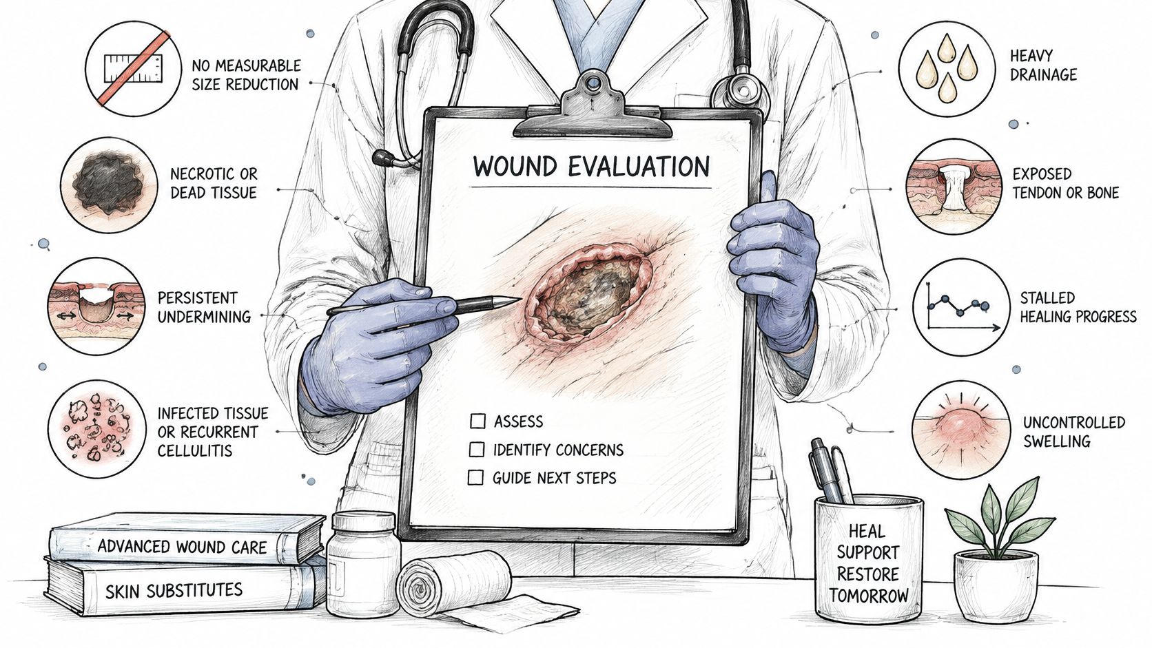
Some findings mean a wound needs more than another covering. We list eight indicators that a wound may deserve surgical evaluation:
- No measurable size reduction: The wound isn't shrinking on serial measurements.
- Necrotic or dead tissue: Nonviable tissue remains in the wound bed.
- Persistent undermining: Tissue separates under the wound edges.
- Infected tissue or recurrent cellulitis: Infection keeps returning.
- Exposed tendon or bone: Deeper structures are visible.
- Persistent slough: Soft, stringy nonviable tissue keeps reforming.
- Recurrent wound breakdown: The wound closes and reopens.
- Minimal improvement: Progress stays poor despite appropriate evidence-based care.
One indicator doesn't mean surgery is needed. Each one flags the need for specialist review through an escalation pathway for surgical wound evaluation.
The checklist below shows how those findings are reviewed in practice.
Why a covering alone cannot fix some findings
Necrotic tissue must be removed before a covering can work. Undermining needs to be opened and cleaned so hidden pockets don't trap drainage and bacteria. Exposed tendon or bone needs structural management, not a covering alone. A heating system that keeps cycling without warming the room is a signal to diagnose the system, not to add more of the same. A stalled wound deserves the same logic.
The video below outlines how a specialist evaluation moves from review to decision.
A specialist review usually starts with history, images, and treatment response, then moves to in-person validation of the wound. The decision may be continued conservative care, a different product, or surgical intervention. At our practice, the surgeon who evaluates the wound is also the one who operates when intervention is appropriate and follows healing afterward. Evaluation can change the level of care, the setting, and the recommendations sent back to your existing team.
Documentation That Decides Coverage and Fit
A skin substitute's name alone does not establish that it fits a wound or qualifies for coverage. The record needs to connect the wound's cause and condition to the product choice, while showing what care has been tried and how the wound responded.
Useful documentation includes:
- Wound cause and serial measurements: Record the wound's cause, size, and depth over time.
- Vascular assessment: Document findings about blood flow to the area.
- Infection status: Record findings at each visit rather than assuming infection is absent.
- Off-loading or compression: Note whether prescribed pressure relief or compression is being used.
- Debridement history: Record what nonviable tissue was removed and when.
- Nutrition and glycemic context: Include relevant factors that affect tissue repair.
- Response to standard care: State what was tried, for how long, and with what result.
- Product rationale: Explain why this product was selected for this wound.
How coverage rules differ from clinical fit
Coverage depends on the payer's written criteria, not just on whether a clinician considers a product appropriate. Medicare guidance on skin substitute coverage focuses primarily on diabetic foot and venous leg ulcers. It generally calls for documentation of conservative treatment and serial wound measurements, along with ongoing compression or other disease-specific care where applicable.
Payer policies often limit treatment duration, the number of applications, and which products may be used on a wound. Check the written policy for the patient's own plan, since requirements can differ.
A clinically reasonable treatment may still be denied, delayed, or stopped if the record does not show the required care or response. Medical necessity and insurance coverage are separate questions. A denial may reflect missing documentation rather than a judgment that the treatment has no clinical value.
More applications or switching products does not automatically improve healing. Direct comparisons between substitute types remain limited, so the record should explain the choice and document the wound's response rather than relying on repeated applications alone.
For example, if a venous wound has complete measurements but no record of compression use, a reviewer may be unable to confirm that standard care was provided. Wound Services reviews history, the wound, imaging, and treatment response virtually, then a surgical wound care nurse validates the wound in person to confirm what was seen. Ask your care team to show how the record supports continuing, stopping, or changing treatment.
Confirm Your Next Step With a Wound Specialist
Start today by gathering the history. Collect prior treatments and their dates, wound photographs, any imaging, and notes on how the wound responded to standard care. Put dates on photographs when you can, because a sequence shows change that a single image can't. This packet saves time and prevents a specialist from repeating questions your record can already answer.
Next, ask your current provider whether specialist review fits your wound. Use these prompts:
- Has the wound shrunk measurably: If not, what explains the stall?
- Are deeper structures involved: Is tendon, bone, or undermining present?
- Has standard care been optimized: Are pressure, infection, and blood flow addressed?
- What is the checkpoint: When will the plan change if there's no response?
What a specialist review looks like
Ask whether a virtual consultation is available. We offer virtual consultations where a wound surgeon reviews the patient's history, images, and the wound by video, usually within days, and we offer a bookable virtual consultation online. That review can happen before you travel, which helps if mobility or distance makes visits difficult.
Expect in-person validation by a wound-care clinician when the video review points to a need for it. Photographs can miss depth, undermining, odor, and tissue quality, so hands-on assessment still matters for many wounds. The escalation pathway connects that assessment to the next level of care when findings warrant it.
Ask for written recommendations to send back to your existing care team, plus a follow-up plan with a defined checkpoint toward closure. Home health nurses, primary care clinicians, and family caregivers all work better from the same written plan. We also offer a way to get wound care now and to contact or refer patients, so referring teams can start that process directly.
Not every wound needs surgery, and not every wound needs a substitute. The right decision rests on documented response. If your wound has stalled, the next step is a specialist opinion rather than another round of the same treatment.
---
If your wound has stopped shrinking, keeps reopening, or hasn't responded to a skin substitute as expected, Comprehensive Wound Services can review your history, images, and treatment response through a virtual consultation, usually within days. Our team coordinates with your existing providers, validates the wound in person when needed, and recommends the level of care your documented response supports. Visit our website to request a consultation or refer a patient for specialist wound evaluation.