The usual advice is to identify a leg ulcer by its appearance, then choose compression for a venous wound or circulation treatment for an arterial wound. That shortcut can be unsafe. A swollen leg with brown skin and a weeping ankle ulcer can still have inadequate arterial blood flow.
The difference between arterial and venous ulcers is primarily a supply problem versus a return-flow problem. The comparison below helps you recognize the clues, but circulation tests determine whether compression, debridement, or vascular intervention is appropriate.
## Table of Contents - Arterial and Venous Ulcers Side by Side - Translating the wound description - Signs that can mislead you - What Drives Each Type of Leg Ulcer - Why the common diagnosis can become a trap - Checking Blood Flow Before You Choose Treatment - The testing sequence - Circulation test results and what they mean - Treatment Choices for Each Ulcer Type in Real Cases - What treatment needs to accomplish - Applying the decision to a patient - How Often Each Type of Leg Ulcer Heals - Reading healing figures without false reassurance - Use the diagnosis to guide reassessment - When a Leg Ulcer Stops Healing - Reassess the cause, not just the dressing - Signs that warrant specialist review
Arterial and Venous Ulcers Side by Side
Arterial vs venous leg ulcers compared
| Feature | Arterial ulcer | Venous ulcer |
|---|---|---|
| Underlying cause | Inadequate arterial blood flow from peripheral arterial disease | Chronic venous hypertension from reflux, obstruction, or both |
| Typical location | Toes, foot, heel, pressure points, anterior lower leg | Gaiter region, especially near the medial malleolus |
| Wound depth and edges | Deep, sharply demarcated or punched out | Shallow, irregular or sloping |
| Wound base | Dry, pale or necrotic, poorly granulating | Granulating, fibrinous, relatively exudative |
| Surrounding skin | Cool, shiny, hairless skin; pallor on elevation; dependent rubor | Edema, hemosiderin staining, lipodermatosclerosis, varicosities |
| Pain pattern | Often painful and may worsen with elevation | Usually less painful; swelling-related discomfort, although substantial pain is possible |
| Pulses and limb temperature | Diminished or absent pedal pulses; cool limb | Pulses usually palpable; limb warm |
| First test to order | Ankle-brachial index and toe pressure | Arterial assessment plus venous duplex ultrasound |
The American Family Physician review of venous ulcers describes these contrasting wound and skin patterns, while emphasizing that clinical appearance requires vascular assessment. The table is a practical lookup, not a diagnostic scorecard. A wound doesn't have to match every feature in either column.
Translating the wound description
The gaiter region is the lower leg around the ankle, where a gaiter would sit. The medial malleolus is the bony prominence on the inside of the ankle. A shallow, draining wound there, surrounded by swelling and brown discoloration, suggests venous disease.
Granulation is the red, developing tissue that helps fill a healing wound. Fibrinous material can appear yellowish, while exudate means wound fluid. Neither yellow tissue nor drainage alone proves infection, and a photograph can't reliably distinguish every type of tissue.
An arterial wound may look almost cut out of the skin, with distinct edges and little healthy tissue developing in its base. Black tissue can represent necrosis, meaning tissue death. A deep wound may expose tendon or fascia, the connective tissue covering underlying structures.
Brown venous staining comes from hemosiderin, an iron-containing pigment left in the skin after blood products escape into surrounding tissue. Lipodermatosclerosis means hardened, inflamed skin and underlying tissue. These changes suggest longstanding venous pressure rather than a recent episode of swelling.
Signs that can mislead you
A red foot isn't necessarily a well-perfused foot. Dependent rubor is redness that appears when the foot hangs down; in arterial disease, the same foot may turn pale when raised. Don't interpret color without considering position, temperature, pulses, and symptoms.
Pain also needs context. Venous ulcers can hurt considerably, particularly when the skin is inflamed or damaged by drainage. Conversely, nerve damage can blunt pain from a serious arterial or pressure-related wound, especially in someone with diabetes.
A palpable pulse is reassuring, but it doesn't measure how much blood reaches the wound. Swelling doesn't establish a venous diagnosis, either. Heart-related fluid retention, medications, immobility, and other problems can cause edema.
Practical rule: Match the wound to the table to decide what to investigate, not what treatment to start.
Mixed disease can produce a combination of rows: a swollen ankle with venous staining, but a cool foot and weak pulses. The wound may be moist rather than dry because venous pressure increases drainage even while arterial supply is limited. Trying to force that presentation into a single visual category can lead to the wrong treatment.
Use the comparison this way:
- Describe the whole limb. Record the wound's location, edges, base, surrounding skin, pain, temperature, and pulses.
- Flag conflicting findings. A venous-looking wound with a cold foot or elevation-related pain needs arterial investigation.
- Request objective testing. An ankle-brachial index and toe pressure assess arterial supply; venous duplex ultrasound investigates reflux or obstruction when venous disease is suspected.
Caregivers can report changes, but pulse assessment and wound-depth evaluation belong with a clinician. Don't probe a wound at home to see whether bone or tendon is exposed.
What Drives Each Type of Leg Ulcer
An ulcer is an open wound that develops when tissue breaks down and doesn't repair normally. Arterial and venous ulcers can both affect the lower limb, but the circulation failures behind them are different.
With venous disease, damaged valves allow blood to move backward, an obstruction interferes with drainage, or both occur together. The resulting pressure encourages fluid leakage and inflammation in the lower leg. Over time, the skin becomes fragile, and minor trauma or spontaneous breakdown can leave an ulcer.
With arterial disease, narrowed or blocked arteries deliver too little oxygenated blood to the tissue. Skin at the toes, heel, or another pressure point may not tolerate ordinary stress. Once injured, it lacks the supply needed to repair itself.
Why the common diagnosis can become a trap
A clinical review of lower-extremity ulcers identifies venous ulcers as 80–90% of all leg ulcers and arterial ulcers as about 10%, while noting that approximately 20% of people with leg ulcers have arterial disease, alone or alongside venous disorders. These figures describe different categories: the proportion classified as arterial ulcers isn't the same as the proportion of patients who have any arterial disease.
Venous ulcers are therefore the more common starting suspicion, but not a safe default diagnosis. An older person with an ankle wound may have chronic venous changes and peripheral arterial disease at the same time. Diabetes, smoking history, kidney disease, and cardiovascular disease make an arterial assessment particularly important.
A common cause can explain the appearance without explaining the whole wound.
The practical distinction is what must be corrected. Venous treatment reduces excessive pressure and improves return flow; arterial treatment aims to improve the blood supply available for tissue survival and repair. A dressing can manage the wound surface, but it can't reopen a blocked artery or correct significant venous reflux.
Our wound conditions guide helps patients recognize the range of problems that can require wound evaluation. Naming the ulcer is useful only when the diagnosis explains both the examination findings and the circulation test results.
Checking Blood Flow Before You Choose Treatment
A leg ulcer's appearance cannot show whether compression is safe. Arterial disease can occur alongside venous dysfunction, so clinicians check blood flow before choosing treatment. The tests answer different questions and must be read together.
The testing sequence
- Examine pulses and measure ABI. The ankle-brachial index compares ankle and arm blood pressure, usually with cuffs and a handheld Doppler. The clinician also checks foot temperature, symptoms, and pedal pulses.
- Add toe measurements when needed. Toe pressure and the toe-brachial index can help when stiff, calcified ankle arteries make ABI unreliable, including in people with diabetes or advanced kidney disease.
- Check venous function. Venous duplex ultrasound assesses blood flow and can identify reflux or obstruction. It does not replace arterial testing before compression.
- Clarify uncertain perfusion. Ankle pressure or transcutaneous oxygen testing, often called TcPO2, can help assess the blood supply available for healing.
Circulation test results and what they mean
The thresholds in this review of venous leg ulcer assessment and treatment algorithms are clinical decision points, not instructions for choosing compression at home.
| Test result | What it suggests | Practical action |
|---|---|---|
| ABI below 0.80 | Supports peripheral arterial disease | Assess arterial status before compression |
| Ankle pressure below approximately 70 mmHg | Associated with delayed wound healing | Review perfusion and the treatment plan |
| ABI around 0.8 or higher | May support standard compression if there are no other contraindications | Consider standard compression after clinical assessment |
| ABI above 0.5 with toe pressure above 50 mmHg | Some published frameworks allow modified compression with arterial disease | Use modified compression only under clinician guidance |
| ABI below 0.4 or toe pressure below 30 mmHg | High ischemic risk | Seek vascular evaluation rather than routine compression |
| Ankle pressure below 50 mmHg, toe pressure below 30 mmHg, or TcPO2 below 25 mmHg | May indicate severe perfusion concerns | Consider urgent imaging and possible revascularization |
| Abnormally high ABI | May reflect a falsely elevated reading from arterial calcification | Add toe pressure, toe-brachial index, or TcPO2 testing |
An ABI from 0.80 to 0.89 supports possible arterial disease, even though it falls within the range where standard compression may be considered. That overlap makes clinical assessment important: the ABI alone does not settle the choice. Symptoms, absolute pressures, wound severity, and test reliability also matter.
A cold foot, rest pain, or worsening tissue loss should prompt further assessment even when ABI appears acceptable. Calcified arteries can resist the pressure of the cuff and produce a falsely reassuring result. When arterial and venous disease coexist, the clinician should document the decision framework and how the limb will be monitored.
Treatment Choices for Each Ulcer Type in Real Cases
Compression is central to venous ulcer treatment, but a venous appearance doesn't clear a limb for tight bandaging. When arterial inflow is severely reduced, compression can worsen ischemia, meaning inadequate blood supply.
This visual comparison highlights the treatment considerations rather than providing clearance for compression.
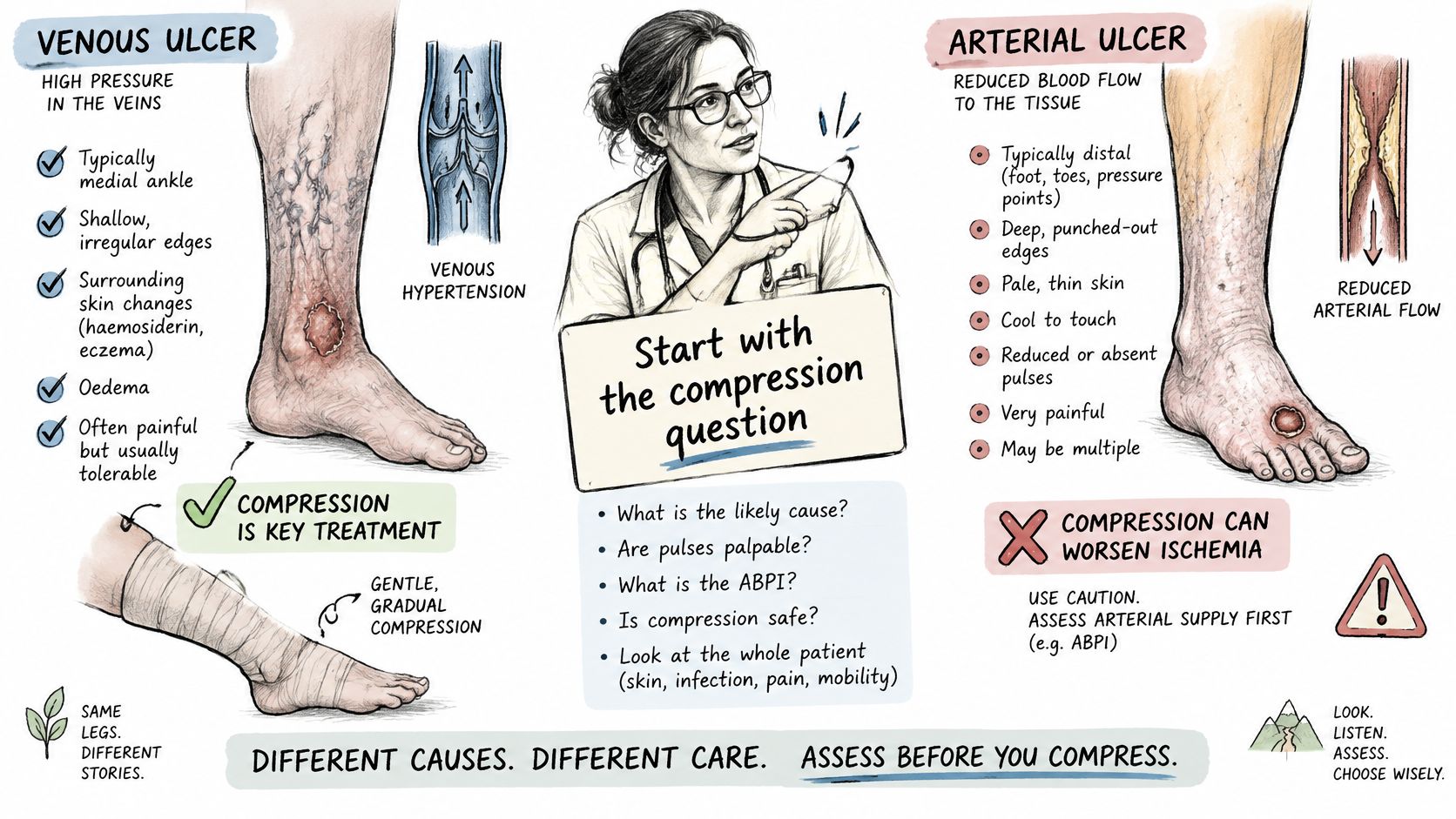
The ACP educational handout on chronic leg wounds notes that about 25% of arterial-ulcer patients also have venous insufficiency, discourages sustained compression when ABI is below 0.6, ankle pressure is below 60 mmHg, or toe pressure is below 30 mmHg, and flags ABPI above 1.2 as potentially requiring further testing. These cautions explain why clinicians don't use swelling alone to select compression.
What treatment needs to accomplish
For a confirmed venous ulcer with adequate arterial supply, treatment usually combines prescribed compression, drainage control, surrounding-skin protection, appropriate elevation, and movement that activates the calf muscles. An absorbent dressing manages exudate; it doesn't replace pressure management. Venous intervention may be considered when duplex findings identify treatable reflux.
For an arterial ulcer, vascular evaluation determines whether blood flow can be improved through revascularization. Pressure offloading protects vulnerable tissue, while medical management addresses contributing cardiovascular risks. Elevation that increases ischemic pain isn't a useful substitute for that assessment.
Debridement requires particular care. Removing dead tissue from a poorly perfused wound can create a larger defect that can't heal. A stable, dry black heel wound generally shouldn't be soaked, softened, or cut away before perfusion and infection have been assessed; wet gangrene or infection requires urgent management.
Treatment priority: Choose dressings for the tissue and drainage, but choose the overall plan for the circulation problem.
The accompanying video offers another way to review arterial and venous wound differences.
Applying the decision to a patient
- Swollen ankle with brown staining and a weeping ulcer. This suggests venous disease. Start with arterial pulse assessment and ABI, adding toe measurements when indicated, then venous duplex; tight compression must wait until arterial safety is established.
- Dry, black heel wound in a person with diabetes. Assess toe pressure alongside ABI because calcification can distort ankle measurements. Offload the heel while arranging evaluation; routine sharp debridement and moisture-based softening of stable eschar must wait.
- Cold foot ulcer with rest pain in an older smoker. This needs urgent vascular assessment, with perfusion testing and imaging as directed. Routine compression and a dressing-only trial must not delay evaluation; sudden coldness, severe pain, numbness, or weakness warrants emergency care.
Our advanced wound care information describes specialty care for complex wounds. Specialty wound treatment should complement, not substitute for, the vascular care an ischemic limb needs.
How Often Each Type of Leg Ulcer Heals
Published healing rates describe groups of patients receiving particular care. They don't predict whether an individual wound will close, and they aren't a reason to postpone treatment.
A systematic review and meta-analysis in the International Wound Journal found a pooled global venous-leg-ulcer prevalence of 0.32% and pooled incidence of 0.17% in its 2023 publication, without a directly comparable pooled arterial-ulcer figure. Prevalence describes how common the condition is; incidence describes new cases. Neither measure tells a patient how likely their current ulcer is to heal.
Reading healing figures without false reassurance
A 2024 cohort study indexed in PubMed reported healing rates of 41% for venous ulcers, 74% for arterial ulcers, and 93% for mixed arterial-venous ulcers in its analyzed cohort. Those results belong to that treated group, not to all patients with those diagnoses.
The higher arterial and mixed rates don't establish that these wounds are easier to heal. Patient selection, wound characteristics, treatment, and follow-up can affect observed outcomes. The figures alone also don't prove that a particular vascular procedure caused the difference.
An ischemic wound still needs a prompt blood-flow assessment. Watching it because a published arterial group had a favorable healing rate would confuse an outcome after care with the safety of withholding care.
Use the diagnosis to guide reassessment
- Venous ulcer remaining open during compression. Review whether the compression is appropriate and consistently used, then reconsider arterial status and whether venous duplex identifies a treatable problem.
- Arterial or mixed ulcer failing to close. Request vascular review of perfusion and whether revascularization is needed or a previous intervention remains effective.
- Diagnosis that doesn't fit the measurements. Reconcile the label with ABI, toe pressure, and duplex findings before changing only the dressing.
A useful healing discussion focuses on your own wound's trajectory: whether its area is decreasing, healthy tissue is developing, drainage is manageable, and surrounding skin is improving. A smaller-looking opening isn't enough if depth or undermining is increasing.
When a Leg Ulcer Stops Healing
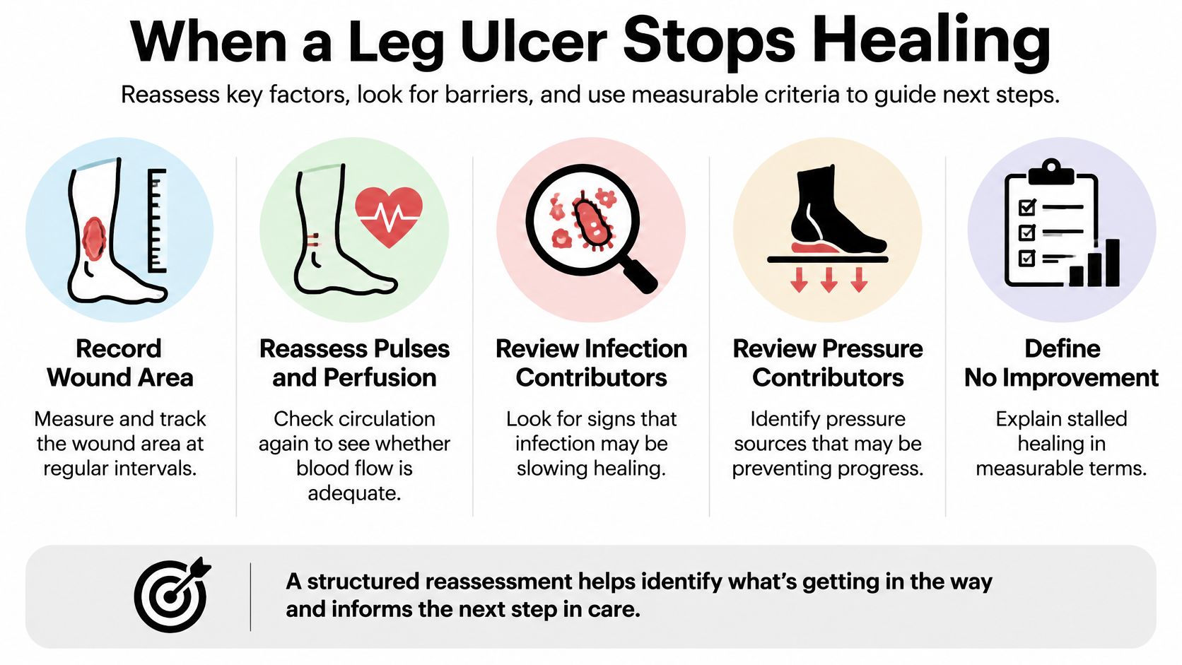
A cleaner-looking dressing does not prove that a leg ulcer is healing. Track its size, depth, tissue, drainage, and surrounding skin to distinguish lasting progress from a temporary surface change.
Use the same measurement method at regular reviews. A clinician can measure length and width with a disposable ruler or estimate area with wound planimetry, which can better account for an irregular shape. Dated photographs taken from a consistent angle with a measurement reference help show change, but do not replace an examination.
Reassess the cause, not just the dressing
A stalled ulcer may reflect missed vascular disease, persistent pressure, infection, or a treatment plan that no longer fits. Recheck pulses and perfusion, inspect footwear and positioning, and assess new redness, warmth, swelling, pain, or drainage. Odor alone does not establish infection.
The 2025 lower-extremity ulceration guidance recommends vascular follow-up at least twice yearly after chronic limb-threatening ischemia or revascularization, with earlier review if a wound stalls or worsens. Repeat blood-flow measurements can assess vessel patency. A deteriorating wound should not wait for a routine appointment.
Signs that warrant specialist review
The surgical wound evaluation escalation pathway identifies eight signs that may warrant evaluation:
- No measurable size reduction. Serial measurements show no decrease.
- Necrotic or dead tissue. Nonviable tissue remains in or around the wound.
- Persistent undermining. Tissue loss extends beneath the visible skin edge.
- Infected tissue or recurrent cellulitis. Infection persists or repeatedly returns.
- Exposed tendon or bone. Deep structures are visible.
- Persistent slough tissue. Adherent slough remains despite appropriate care.
- Recurrent wound breakdown. The wound repeatedly reopens.
- Minimal improvement despite appropriate evidence-based care. The response to treatment remains limited.
Bring wound measurements, dated photographs, a current medication list, previous ABI or toe-pressure results, imaging reports, and a record of treatments used. Describe what has changed and whether pain occurs at rest, with elevation, or during walking.
These findings warrant specialist reassessment. Surgical evaluation does not automatically lead to surgery. Conservative care remains the foundation, with intervention considered only when clinically appropriate.
Wound Services offers virtual and in-person specialty evaluation for complex, stalled, and potentially surgical wounds, coordinated with existing providers. If a leg ulcer is not shrinking or its circulation-related diagnosis remains uncertain, arrange a wound evaluation with Comprehensive Wound Services to review the appropriate next step.