Yellow slough is devitalized tissue in a wound bed, but it doesn't by itself mean the wound is infected or that you should remove it at home. The color is a clue to assess alongside the wound's drainage, surrounding skin, pain, and healing progress, not a diagnosis on its own.
A caregiver lifts a dressing and finds a new yellow layer. It's understandable to worry that antibiotics are needed that night, but yellow tissue alone can't answer that question. What matters is what the tissue looks like and whether the wound has other concerning changes.
## Table of Contents - Yellow on a Wound Is Not a Diagnosis - What you can observe safely - How Yellow Slough Differs From Other Wound Tissue - What to Check Besides the Yellow Color - Why Yellow Slough Returns After Cleaning - Reassess the wound, not just the surface - Care That Matches the Slough You See - Common Wound Scenes and What They Suggest - When Persistent Slough Needs a Specialist
Yellow on a Wound Is Not a Diagnosis
Yellow is a poor stand-alone triage signal. Slough is nonviable material in the wound bed, made up of accumulated dead cellular debris that may also contain fibrin, bacteria, and inflammatory by-products. It can be loose or firmly attached, thin and stringy or thick and waxy, wet or dry. Its appearance doesn't tell you by itself whether the wound is infected.
The wound management guidance from East London NHS Foundation Trust distinguishes slough from clinical infection. Infection is judged using additional signs, such as spreading redness, increasing pain, warmth, swelling, pus-like drainage, a new bad odor, feeling unwell, or healing that has stalled. A swab isn't automatically needed just because yellow tissue is visible; a clinician decides whether sampling is appropriate when infection is suspected.
Clinicians document the type of tissue and the proportion of the wound bed it covers, rather than treating “yellow” as a diagnosis. Slough can delay healing because it occupies the wound bed and can support bacterial growth, making it harder for healthy granulation tissue and new skin to develop. But a wound that's gradually shrinking, with stable drainage and calm surrounding skin, is different from one with a sudden increase in yellow material, pus, spreading redness, warmth, swelling, or pain that seems out of proportion.
What you can observe safely
At home, note changes in the wound's size, the amount and character of drainage, any change in odor, the skin around the wound, pain, and whether the person feels systemically unwell. These observations help a clinician judge the whole picture. Yellow or brown material can also warrant assessment for a fistula or another underlying problem, rather than routine slough care.
Don't scrape, cut, or pull adherent slough. That can injure viable tissue or cause bleeding, and it's especially risky on the foot or a limb with arterial disease. Get specialist advice before any debridement in those situations.
How Yellow Slough Differs From Other Wound Tissue
The same color can describe different wound findings. Use this comparison as a starting point, not as a way to diagnose infection by sight.
| Finding | Usual look | What it often means | Proves infection alone |
|---|---|---|---|
| Yellow slough | Yellow, tan, or cream; stringy and wet or thick and waxy | Devitalized debris that can block granulation | No |
| Healthy granulation | Beefy red, moist, and bumpy | New tissue filling the wound | No |
| Pale or dusky granulation | Pink, gray, or purple and easily damaged | Possible poor perfusion, edema, or bioburden stress | No |
| Fibrinous film | Thin yellow coating over a viable bed | Fibrin from exudate; not always thick slough | No |
| Eschar | Black or brown, dry, and leathery | Necrosis; stable ischemic eschar isn't a home-softening task | No |
| Purulent drainage | Thick yellow, green, or cloudy fluid | Possible infection when other signs are present | No; it raises suspicion |
| Exposed tendon or bone | Shiny cord or hard pale structure in the base | Wound depth beyond skin; needs specialist review | No |
The distinctions matter because a thin film isn't the same as a thick layer of adherent slough, and fluid isn't the same as tissue. A small amount of yellow fibrin over an otherwise viable wound bed doesn't automatically signal infection. Likewise, red granulation tissue is often part of healing, while pale or dusky tissue can prompt a clinician to consider whether circulation, swelling, or other wound stresses are affecting the bed.
Pus-like drainage deserves attention, especially when it appears with spreading redness, warmth, swelling, worsening pain, odor, or systemic symptoms. But drainage color alone still doesn't establish the diagnosis. A shiny tendon or visible bone is a different kind of warning: it suggests the wound extends beyond the skin and needs specialist assessment.
Think of slough as rubble left on a building site, granulation as the new framework, and pus as a leak that needs its cause found, not another coat of paint. When describing what you see, estimate what percentage of the visible bed is yellow slough versus red tissue. That tissue mix is more useful than the single word “yellow.”
What to Check Besides the Yellow Color
Use the same order each time you check a wound. A consistent record helps a clinician see a trend instead of relying on one photograph or one description.
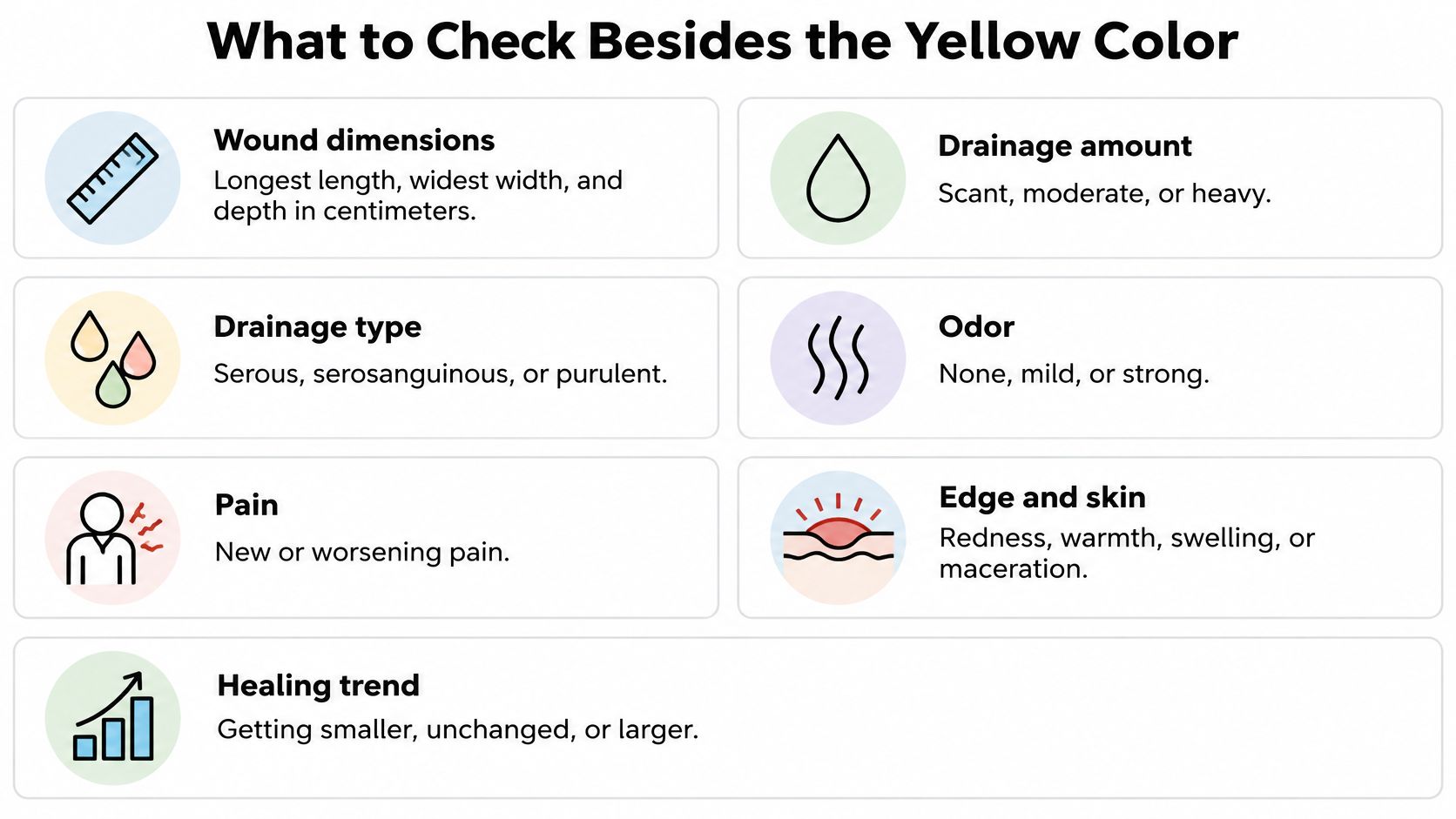
- Measure the wound. Record its longest length, widest width, and depth in centimeters. Estimate the percentage of the bed that's slough, red tissue, or eschar. If you can't measure depth safely, don't probe the wound; report that it wasn't measured.
- Check for undermining or tunneling. A small opening at the surface can hide a pocket or tract beneath it. Tell the clinician if the wound edge seems to overhang or if a deeper area is already known.
- Describe drainage. Note whether it's scant, moderate, or heavy, and whether it's clear and watery, blood-tinged, or cloudy and pus-like. The Royal Cornwall Hospitals NHS Trust wound care guideline recommends assessing wound dimensions, undermining, exudate, odor, surrounding inflammation, pain, and systemic signs rather than relying on color alone. It also notes that persistent slough can delay progression toward granulation and new skin.
- Record odor as a change. Note whether there's no odor, a mild odor, or a strong odor, and whether it's new or different from the wound's usual smell.
- Look at the surrounding skin. Record whether it's intact or softened and white from moisture, and whether redness, warmth, or swelling is spreading away from the wound edge.
- Describe pain. Note pain at rest and during dressing changes. Say whether it's new, worsening, or disproportionate to what you'd expect.
- Check how the person feels. Record fever, chills, rising blood glucose in someone with diabetes, confusion, or feeling markedly unwell. These findings can change the urgency of an assessment.
A newly increasing slough burden, malodor, purulence, spreading redness, disproportionate pain, or stalled healing warrants clinician review.
A wound swab isn't an automatic response to visible slough. Clinicians consider sampling when the overall picture suggests clinical infection. To make follow-up more useful, keep a brief log at each dressing change: measurements, tissue mix, drainage, odor, surrounding skin, pain, and how the person feels. For more on the kinds of wounds that may need assessment, see common wound conditions.
Why Yellow Slough Returns After Cleaning
A wound can look cleaner after a visit and then show yellow material again days later. That doesn't necessarily mean the caregiver cleaned badly. Cleaning can remove visible debris without addressing the conditions that allow new debris to accumulate.
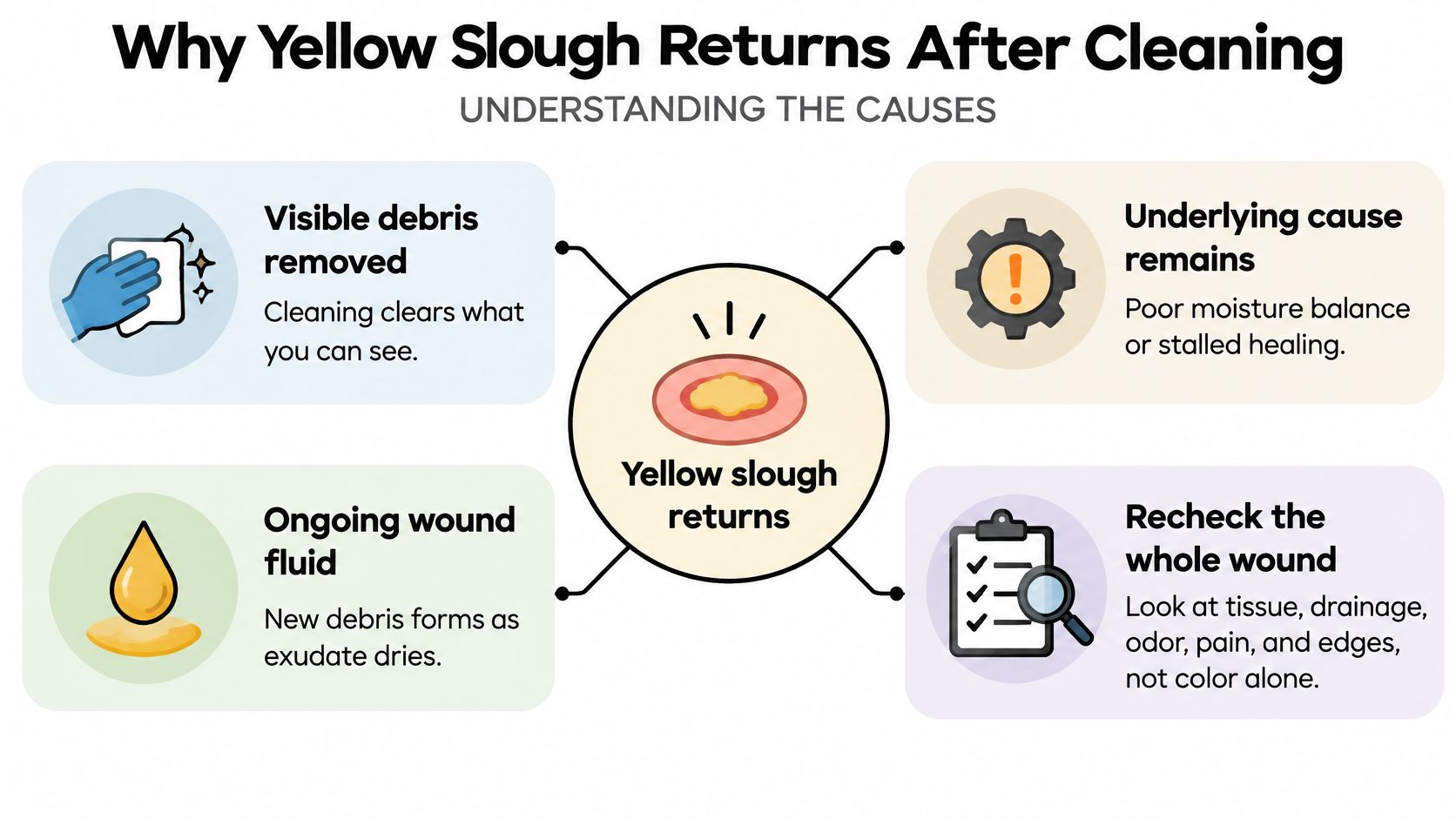
A clinician may consider biofilm, a community of microbes that can re-form after disruption. The expert literature on wound biofilm and stalled healing identifies impaired healing over several weeks, recurrent or excessive drainage, rapid slough re-accumulation after debridement, and subtle inflammation without obvious infection as reasons to suspect biofilm involvement. That possibility calls for reassessment, not blame or more forceful cleaning.
Other factors can keep the cycle going. A dressing may not manage the amount of fluid, pressure or shear may persist at a heel or sacrum, and poor arterial supply or edema can compromise the wound bed. Ongoing inflammation or an unrecognized deeper pocket, tract, or exposed structure may also be part of the problem.
Reassess the wound, not just the surface
Compare size and depth over time, and record drainage and odor. Check whether pressure is being relieved, whether circulation has been assessed, and whether diabetes control or nutrition needs attention. If yellow tissue returns rapidly after appropriate care, the treatment plan or the explanation for the wound may need review.
Debridement removes devitalized tissue, and clinicians may need to repeat it when slough or biofilm returns. It doesn't diagnose why the wound is stalled or replace treatment of its cause. The building-site comparison applies here: the rubble may return if a leak or unstable ground remains unaddressed.
A clinician should review persistent undermining, exposed tendon or bone, recurrent infection, or little improvement despite appropriate care. If you're tracking progress, compare dated measurements and photos rather than judging the wound by whether it looks clean immediately after a dressing change.
Care That Matches the Slough You See
Treatment should fit the slough's adherence, the amount of drainage, circulation, pain, infection status, and the person's overall risk. The 2024 wound debridement consensus document describes cleansing as a way to remove surface contaminants and loose debris, while debridement removes devitalized or adherent tissue. The right approach isn't determined by the word “yellow.”
| Pattern | Usual first approach | Escalate when |
|---|---|---|
| Dry or low-exudate adherent slough | Moisture-retentive dressing or hydrogel to support slow autolysis | Ischemia, rising pain, or no softening |
| Moderate to high exudate | Absorptive hydrofiber or similar dressing to balance moisture | Maceration, rapid refill, or new odor |
| Loose surface debris only | Gentle cleansing of contaminants | Cleaning causes bleeding or new pain |
| Large, firmly adherent burden | Clinician debridement, which may be enzymatic, conservative sharp, or surgical | Foot wound, arterial disease, anticoagulation, or uncertain depth |
| Slough back within days of proper care | Reassess biofilm, pressure, perfusion, and the wound's cause; a clinician may repeat debridement if indicated | Little size change over several weeks |
For dry or low-exudate slough, moisture-retentive dressings can support autolysis: the body's own enzymes soften and break down the tissue. Hydrogels are generally suited to this setting because they rehydrate the wound bed, but autolysis can be slow when the burden is large. Don't let a slow trial delay review if infection or deep involvement is suspected.
When drainage is moderate to heavy, an absorptive hydrofiber or related dressing may be more appropriate to manage fluid and protect the surrounding skin. Loose debris can be addressed with gentle cleansing, but cleaning isn't permission to scrape the wound base.
Sharp or operative debridement belongs to appropriately trained clinicians. They need to be especially cautious with ischemia, anticoagulant use, neuropathy, exposed vital structures, or uncertain wound depth. Debridement may disrupt biofilm defenses and improve access for treatment, but it doesn't replace pressure relief, circulation assessment, infection control, moisture balance, or care of the underlying wound cause. If you need information about specialist options, see advanced wound care services.
Common Wound Scenes and What They Suggest
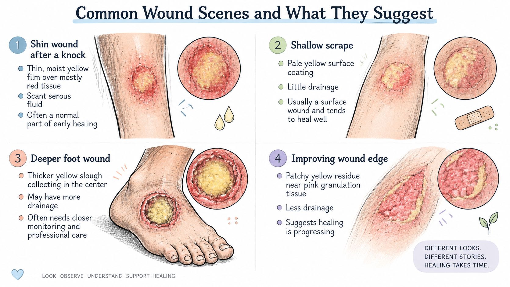
A shin wound after a knock has a thin, moist yellow film over mostly red tissue. There's only a little clear drainage, the surrounding skin looks calm, and the wound is a little smaller after a period of follow-up. That pattern doesn't call for antibiotics based on color alone; follow the existing care plan and keep monitoring. Photograph the wound and report the estimated yellow percentage, drainage, pain, and whether its size is improving.
A person who sits for long stretches has a sacral pressure injury. Thick yellow slough returns after each cleaning, drainage soaks the dressing, and the measurements barely change. Harder scrubbing isn't the answer. The wound needs review of pressure relief and the stalled healing, along with a reassessment of drainage management. Send a dated image and tell the clinician how quickly the dressing becomes wet, whether pain has changed, and whether the wound size has shifted.
A person with diabetes has a foot ulcer with increasing yellow-gray material, a new odor, and pain or a finding that suggests deeper tissue involvement. This calls for urgent same-day clinical assessment. Don't debride it at home before the wound's depth and circulation are assessed. Share a photograph only if doing so won't delay care, and describe the odor, drainage, pain, and change in size.
A surgical wound had been closing, then begins leaking yellow or brown fluid from one point rather than showing a broad film across the bed. That pattern may need assessment for a fistula or another underlying problem, not a label of ordinary slough. Take a dated photo and tell the clinician where the fluid appears to come from, how much there is, whether pain has changed, and whether the wound had been getting smaller.
When Persistent Slough Needs a Specialist
Seek urgent local care for spreading redness, fever, rapidly worsening pain, pus accompanied by systemic symptoms, a wound that's blackening, or any glimpse of tendon or bone. Those signs need timely clinical assessment, not a home attempt to remove tissue.
A specialty wound review is also appropriate when slough persists alongside no measurable reduction in wound size, necrotic tissue, persistent undermining, infected tissue or recurrent cellulitis, exposed tendon or bone, recurrent wound breakdown, or minimal improvement despite appropriate evidence-based care. These are eight indicators CWS lists for possible surgical evaluation. A review isn't an automatic operation. It's a chance to reassess the wound's history, images, circulation, and response to treatment.
For people in the Washington, DC area with a complex, stalled, or potentially surgical wound, Wound Services offers virtual consultations in which a wound surgeon reviews the history, images, and wound by video, usually within days. In-person assessment is also available, and surgery is considered only when clinically appropriate. CWS reports that approximately 18,521 wounds have improved or healed; individual results vary with wound type, health history, and care plan. To understand the possible next steps, review the surgical wound evaluation escalation pathway.
While arranging review, gather dated photographs, measurements, a list of dressings already tried, and notes about drainage, odor, pain, and changes in the surrounding skin. Don't attempt to remove adherent slough while waiting.
---
If yellow tissue keeps returning, the wound isn't shrinking, or you're concerned about deeper involvement, the clinic offers virtual and in-person specialty evaluation for complex, stalled, and surgically appropriate wounds. Share your wound history and available images with Comprehensive Wound Services to explore whether a specialist review is a suitable next step.