
You've been changing dressings, keeping appointments, and trying to protect a wound that still won't close. Now someone has mentioned wound flap surgery, and you're wondering whether it means a bigger operation, a better chance of healing, or more care than you can manage at home.
A flap can help repair a wound that simpler treatments can't adequately cover. But deciding whether it's appropriate means looking beyond the operation, including your health, the cause of the wound, and who will help protect the repair afterward.
## Table of Contents - What Wound Flap Surgery Is and How a Flap Works - Common flap types and what they carry - Why Surgeons Choose a Flap Over Simple Closure - Benefits and trade-offs - How Surgeons Decide Whether You Are a Candidate - From medical history to a treatment decision - Candidacy risk factors and what teams plan - What Success Rates Look Like and Where Flaps Fail - Recurrence at 12 months by ulcer site - How a repair can fail - Recovery at Home and in Skilled Facilities - A discharge-readiness checklist - Warning Signs That Need Urgent Attention - Make reporting straightforward - Get a Specialist Evaluation Before Deciding on Surgery
What Wound Flap Surgery Is and How a Flap Works
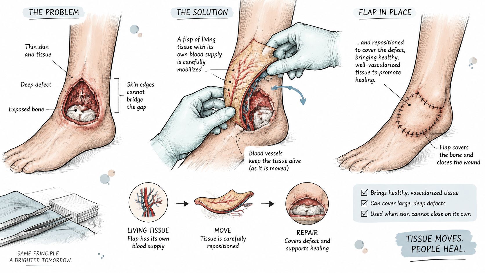
Consider a deep wound with thin surrounding skin and exposed bone. Stitches might pull the edges together, but they can't replace the missing tissue underneath.
Wound flap surgery moves healthy tissue into that defect with its blood-vessel network. For a flap that stays attached, think of moving a house while keeping its plumbing connected, rather than carrying over the furniture and rebuilding. With a free flap, the surgeon disconnects and then reconnects that plumbing at the new location.
The diagram illustrates how living tissue can fill and cover a deep wound.
Common flap types and what they carry
Surgeons consider three connected parts: the tissue being moved, the vessels supplying and draining it, and the wound bed receiving it.
| Flap type | Tissue carried | Reported recurrence (pressure ulcers) |
|---|---|---|
| Fasciocutaneous | Skin and underlying fat, with supporting fascia | 8.0% |
| Perforator-based | Skin and fat supplied by an identified perforating vessel | 7.5% |
| Musculocutaneous | Muscle with overlying skin | 6.3% |
| Free flap | Tissue lifted completely and reconnected to new vessels under a microscope | No pressure-ulcer recurrence figure available in the cited comparison |
The pressure-ulcer recurrence figures in this table come from a systematic review discussed in an International Wound Journal article: 8.0% for fasciocutaneous, 7.5% for perforator-based, and 6.3% for musculocutaneous flaps. They aren't a ranking of which operation is best for you.
The first three names describe tissue composition or blood supply; free flap describes how tissue is transferred. These categories can overlap.
A head-and-neck free-flap series reported 97.6% overall flap survival, illustrating what microsurgical transfer can achieve in that setting, not predicting every wound patient's outcome.
A flap is living tissue, not a patch. Its circulation must work for the repair to survive.
Why Surgeons Choose a Flap Over Simple Closure
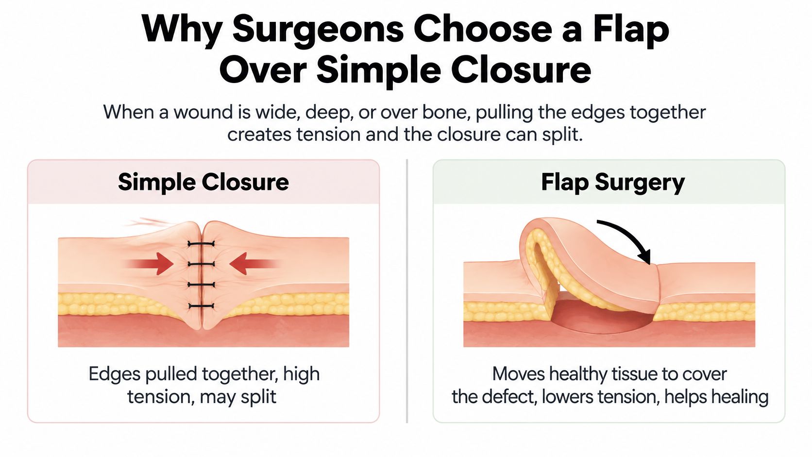
When a wound is wide, deep, or over bone, pulling the edges together creates tension and the closure can split. Even if the surface closes, an unfilled cavity may remain underneath.
This comparison shows the difference between pulling existing edges together and bringing additional tissue into the defect.
Benefits and trade-offs
| Potential benefit of a flap | Trade-off to consider |
|---|---|
| Brings tissue with its own circulation | The flap can lose blood flow |
| Fills a deep cavity, called dead space | Creating and positioning the flap requires more surgery |
| Adds padding over bone, tendon, or a joint | The donor site also needs to heal |
| Covers selected areas unsuitable for a skin graft | Recovery may involve substantial positioning and mobility restrictions |
A skin graft transfers skin without its own connected blood supply and must obtain nourishment from the receiving wound bed. A flap can supply tissue volume and circulation where a thin covering wouldn't address the underlying defect.
Flap reconstruction may require a separate operation after wound preparation, and it generally involves more operative work than straightforward stitching. Some patients need further procedures if the tissue breaks down or the repair needs adjustment.
For example, someone with a deep pressure wound over the tailbone may need tissue to fill the cavity and protect the bone. If that person returns to prolonged sitting without an appropriate cushion, turning schedule, and seating assessment, the repaired area remains vulnerable.
A flap isn't an automatic upgrade. Surgeons choose it according to depth, location, available tissue, and the limitations of simpler options. Dressings, pressure relief, infection management, and other forms of advanced wound care may remain appropriate.
Ask your surgeon: What would happen to this wound if we only stitched it closed?
How Surgeons Decide Whether You Are a Candidate
A surgical evaluation should explain both whether the wound needs reconstruction and whether you can safely complete recovery. The decision follows a sequence rather than a single photograph or measurement.
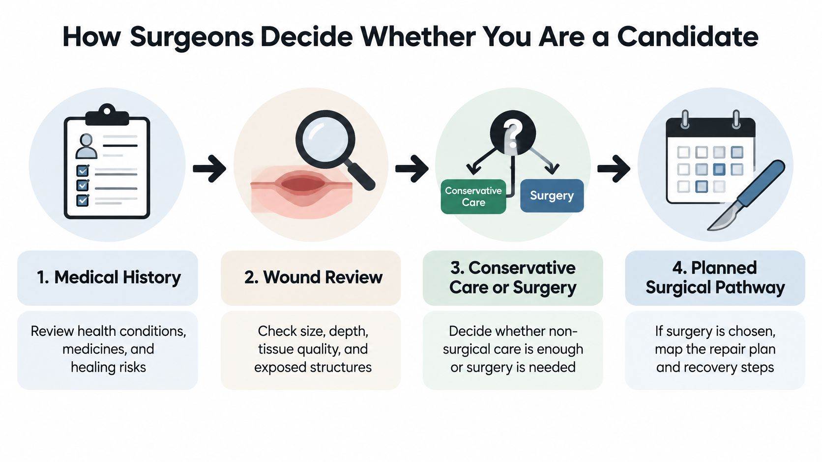
From medical history to a treatment decision
- Review the medical history. The team checks diabetes, vascular disease, immune status, previous operations at the site, blood-thinning medicines, and mobility. These findings help identify anesthesia risks and barriers to healing.
- Examine the wound. The clinician assesses size, depth, tissue quality, infection, exposed structures, and undermining, where damage extends beneath the skin edge. Imaging may help answer questions about deeper structures.
- Choose conservative care or surgery. The surgeon considers whether better pressure relief, wound preparation, infection treatment, or other nonsurgical care could allow progress.
- Plan the surgical pathway if appropriate. This includes the proposed repair, preparation needed beforehand, and arrangements for protecting it afterward.
At Wound Services, we review the history, wound, imaging, and treatment response virtually. A surgical wound care nurse then validates the wound in person to confirm what was seen.
Our evaluation indicators include:
- Stalled size: No measurable reduction.
- Dead tissue: Necrotic tissue remains.
- Persistent undermining: Pockets continue beneath the edges.
- Infection: Infected tissue or recurrent cellulitis.
- Exposed structures: Visible tendon or bone.
- Persistent slough: Adherent nonviable tissue remains.
- Repeated breakdown: The wound closes and reopens.
- Limited response: Minimal improvement despite appropriate evidence-based care.
These findings justify assessment; they don't establish that a flap is necessary.
Candidacy risk factors and what teams plan
A large pressure-ulcer flap outcomes analysis identified low body mass index, active smoking, and ischial location as independent recurrence risk factors.
| Factor | Documented effect | What the team plans |
|---|---|---|
| Low body mass index | Independently increased recurrence risk | Nutrition assessment and optimization before surgery |
| Active smoking | Independently increased recurrence risk | Cessation plan timed before the operation |
| Ischial tuberosity location | Independently increased recurrence risk | Pressure redistribution and seating plan |
Low weight should prompt a closer nutrition assessment, not an assumption that gaining weight solves the problem. Likewise, smoking cessation needs practical support, and a seating plan must fit the person's actual transfers and daily activities.
A 2023 pressure-ulcer reconstruction cohort reported 90% initial flap success, but 16% recurrence among 105 patients assessed after 12 months. Those are different outcomes, reinforcing why candidacy includes the whole person and the recovery environment.
What Success Rates Look Like and Where Flaps Fail
Initial flap survival, wound healing, and freedom from recurrence aren't interchangeable. A flap can keep healthy blood flow while one edge of the incision pulls apart. A wound can close, hold for months, and then reopen under the same pressure that caused it. Each of these is a different endpoint, and different studies count different ones, which is why a single "success rate" rarely tells the whole story.
Recurrence at 12 months by ulcer site
A 2023 pressure-ulcer reconstruction cohort reported the following site-specific recurrence figures at 12 months, among patients available for longer-term assessment.
| Ulcer site | Recurrence at 12 months |
|---|---|
| Sacral | 18% |
| Trochanteric | 35% |
| Ischial tuberosity | 47% |
| All sites combined (17 of 105 patients) | 16% |
The ischial tuberosities, the sitting bones beneath the buttocks, take repeated loading every time a person sits. That mechanical exposure helps explain why this site recurred most often in the cohort, although location alone doesn't predict any one person's outcome.
Sacral wounds, near the lower spine, recurred least often here. That still doesn't make them low-risk wounds, and pressure redistribution remains part of the plan for every site.
How a repair can fail
- Blood-flow failure: Inadequate inflow or blocked venous drainage can cause tissue death, called necrosis.
- Edge separation: The incision can open, known as dehiscence, even when much of the flap remains alive.
- Infection beneath the repair: Fluid or infected tissue may require drainage, antibiotics, or further surgery.
- Recurrent ulceration: The repaired area can break down again under continued pressure or shear.
These failures also surface on different timelines. Circulation problems can appear immediately after surgery, while recurrent ulceration may take months of ordinary sitting and transfers to develop. Studies that stop counting early will look better than studies that follow patients for a full year.
A 2025 groin-flap reconstruction study reported 5.4% partial flap necrosis alongside 98.9% flap survival at 12 months. Partial tissue loss and overall survival can coexist in the same result.
A 2025 review of perforator flaps for pelvic-trochanteric pressure ulcers, covering 4,105 patients, reported 23.9% overall complications and 12.4% recurrence. These figures describe that review's population, not a universal forecast for flap surgery.
Before accepting a success percentage, ask what was measured: living tissue, a closed wound, avoidance of another operation, or healing without recurrence.
Also ask what the team does if circulation becomes compromised immediately after surgery. Monitoring and access to urgent reassessment matter because some problems require prompt surgical treatment, not a routine dressing change. Closing the tissue is one stage; the flap still has to survive the months of pressure, transfers, and daily care that follow.
Recovery at Home and in Skilled Facilities
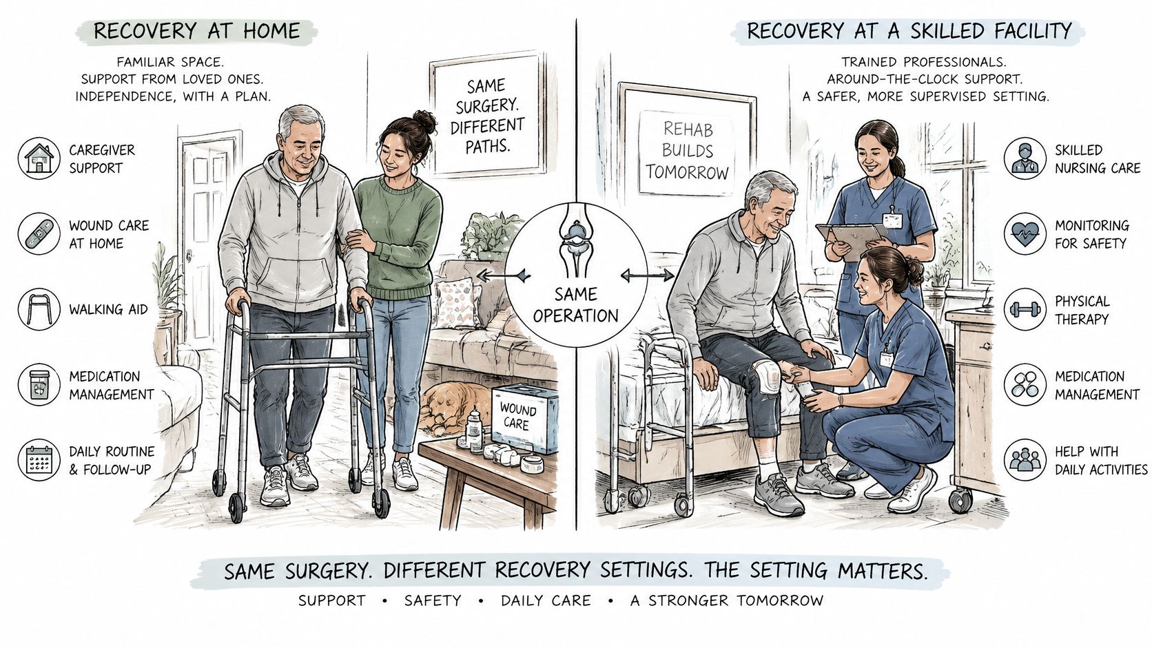
A flap that survives the operating room still has weeks of healing ahead. The surgery closes the tissue, but the discharge setting is where that closure either holds or fails. Two patients can leave with nearly identical repairs and need very different plans: one may go home with a trained caregiver, suitable equipment, and home-health support, while another needs the nursing and transfer assistance that a skilled facility provides.
A fresh flap works like a newly transplanted plant. It survives the move only if the conditions afterward, pressure kept off the area, steady circulation, careful handling with every turn and transfer, stay right day after day. A home can meet those conditions, and so can a facility. The illustration contrasts the two environments.
Recovery can also take longer than families expect. In a study involving patients with spinal cord injury, 83.7% were healed one year after surgery, while 44.2% required a return to the operating room. Eventual healing does not necessarily mean an uncomplicated recovery, and these findings should not be generalized to every flap patient.
Neither home nor a facility is automatically the better choice. The practical question is whether the setting can reliably deliver the prescribed care, including nights, weekends, and transport to appointments.
A discharge-readiness checklist
- A reliable caregiver: Identify who helps with positioning, hygiene, and monitoring, including backup coverage.
- Appropriate equipment: Confirm the bed surface, wheelchair cushion, and transfer equipment match the restrictions.
- Dressing support: Name who changes dressings, how they are trained, and how supplies arrive.
- Transportation: Arrange a way to attend appointments without violating positioning instructions.
- Meal support: Plan adequate food, fluids, and any individualized nutrition recommendations.
- Medication instructions: Keep an updated list, including clear directions for blood thinners. Do not stop or restart them independently.
- Written escalation instructions: Record warning signs, contact numbers, and where to go urgently.
Before discharge, the surgical team should write down the pressure restrictions, the turning schedule, the permitted transfers, and how activity will progress over time. There is no single safe sitting schedule that fits every flap, so the written plan matters more than a general rule.
For patients discharged to a nursing facility, virtual wound-care coordination in skilled nursing can help facility staff and the surgical team stay aligned on the care plan. At Wound Services, follow-up and recommendations return to your existing care team, and virtual follow-up tracks the wound to closure.
This video is an additional resource to discuss with your care team, not a substitute for individualized instructions.
The safest discharge plan is agreed, assigned, and written before you leave. Closing the wound is one stage of the pathway. The setting you go home to shapes the rest of it.
Warning Signs That Need Urgent Attention
| Sign | What it may indicate | Action |
|---|---|---|
| Flap turns dark, purple, or cold | Compromised blood flow | Emergency care now |
| Redness, warmth, or swelling spreading past the wound edge | Possible infection | Same-day contact with the team |
| Cloudy, foul-smelling, or increasing drainage | Possible infection | Same-day contact with the team |
| Gap opening along the incision | Wound dehiscence | Prompt review; keep the area covered |
| Fever or chills | Possible infection | Urgent contact with the team |
| Pain that grows rather than eases | Possible complication | Call the team for review |
Some tenderness and swelling can occur during early recovery. What deserves attention is a change from the expected pattern, especially altered color or temperature, worsening drainage, spreading inflammation, or separation of the closure.
Don't assume increasing pain means you need more pain medicine. Contact the team so they can decide whether the wound needs examination.
Make reporting straightforward
A compromised flap is time-sensitive. If it becomes cold or changes markedly in color, follow the emergency pathway immediately and notify the surgical team; don't wait for a scheduled appointment or a reply to an uploaded photograph.
Keep an opening incision covered with the prescribed dressing. Don't probe beneath the flap, pull at stitches, or apply heat or ice unless your surgeon has specifically instructed you to.
- Photograph consistently: Follow the team's schedule, using similar lighting and angles without disturbing protected dressings.
- Keep a short log: Record pain changes, drainage, odor, and any new symptoms.
- Post the emergency number: Put daytime and after-hours contacts on the refrigerator and in your phone.
A photograph can help explain a change, but it shouldn't delay urgent care.
If fever accompanies confusion, faintness, breathing difficulty, or rapid deterioration, seek emergency medical help rather than waiting for a routine callback.
Get a Specialist Evaluation Before Deciding on Surgery
A useful consultation should leave you with a clear recommendation and the reasons behind it, even if the recommendation is to continue nonsurgical treatment. Bring enough information to distinguish a wound that needs different conservative care from one that may need reconstruction.
- Gather the records. Bring your medical history, current medication list, dated wound photographs, imaging reports, and previous procedure notes if available. Include allergies and the names of clinicians currently managing the wound.
- Document what's been tried. List dressings, pressure-relief equipment, infection treatment, and other therapies, including how long they were used and whether measurements improved. Explain any practical difficulties following the plan.
- Identify the evaluation indicators. Ask which findings apply to your wound and what each means. Exposed bone, for example, raises different questions from a shallow wound that has stopped shrinking.
- Clarify who evaluates and treats you. Our surgical wound evaluation pathway connects assessment with intervention when appropriate. At CWS, the surgeon who evaluates the wound is the surgeon who operates when intervention is appropriate and follows the healing afterward.
- Request a realistic recovery plan. Ask about the expected healing timeline, positioning restrictions, follow-up schedule, and possible need for further treatment. Use the discharge-readiness checklist to confirm who owns each task.
- Confirm the recommendation with your existing clinicians. Make sure the wound-care provider, primary clinician, home-health team, or facility receives the plan. Ask how conflicting instructions will be resolved.
Confirm locally which virtual or in-person review formats are available before booking. An evaluation doesn't commit you to surgery, and conservative care may remain the right path.
The decision should weigh the wound, your overall health, and the support available where you'll recover. Closing tissue is an important step, but the larger aim is a repair you can protect and maintain.
---
Specialty evaluation is offered for complex, stalled, and potentially surgical wounds, with coordinated treatment when intervention is appropriate. Visit Comprehensive Wound Services to ask about evaluation options and bring your wound history, medication list, and recovery-support questions to the consultation.