Many diabetes-related foot ulcers heal, but a substantial share do not. A wound that stays open may need more than a different dressing. Clinicians also assess blood supply, nerve function and pressure on the foot, since each can affect healing.
For patients and families, the useful question is: “What is preventing it from closing?” Understanding the possible barriers helps you know when routine follow-up may be appropriate and when to contact the care team for reassessment.
## Table of Contents - Warning Signs That a Foot Wound Is Not Healing - Signs that change how quickly to act - Keep a useful record - Underlying Causes Behind a Stalled Foot Wound - Different causes need different responses - Match the questions to your history - Circulation and Nerve Tests Clinicians Use - What each test adds - When results don't agree - How Clinicians Assess the Wound Step by Step - From history to a working explanation - What meaningful progress looks like - Treatment Choices and When Surgery Is Considered - Pressure relief and local wound care - Infection, blood flow and surgical decisions - Getting a Specialist Review and Acting This Week - What a specialist review can clarify - A practical plan for this week
Warning Signs That a Foot Wound Is Not Healing
A wound that isn't measurably improving needs reassessment, not an indefinite repeat of the same dressing. Slow healing and stalled healing aren't identical. A wound may remain open while gradually becoming smaller and shallower, but an unchanged, enlarging or repeatedly reopening wound suggests that something still needs attention.
Healing also doesn't erase the underlying risk. The IWGDF prevention guideline reports that diabetes-related foot ulcers recur in approximately 40% of people within one year after healing and 65% within three years. Continued protection and follow-up matter even after the skin closes.
Signs that change how quickly to act
Use the following signs to choose the urgency of care, not to diagnose the problem yourself.
| Warning sign | What it may mean | Act within |
|---|---|---|
| No measurable size reduction by the planned review | Treatment isn't working; circulation or pressure may need reassessment | Contact the treating team promptly for an earlier review |
| Wound remains open beyond the expected course or repeatedly reopens | Chronic or recurrent tissue breakdown | Arrange a prompt reassessment |
| Spreading redness, warmth or swelling | Cellulitis or spreading infection | Same day |
| Foul odour, pus or cloudy drainage | Infection, possibly involving deeper tissue | Same day |
| Black, grey or dead tissue | Tissue death, possibly related to poor blood flow | Same day |
| Exposed tendon or bone | Deep tissue involvement, possible bone infection | Urgent, same day |
| Cold, pale or blue foot, or pain at rest | Inadequate arterial blood supply | Urgent assessment; emergency care now if sudden or worsening |
| Fever, chills or confusion with a foot wound | Possible systemic infection | Emergency care now |
A suddenly cold or discoloured foot is particularly time-sensitive. Don't wait for a routine appointment if the change is new, especially with severe pain, weakness or increasing numbness.
Pain isn't a reliable safety signal. Nerve damage can reduce sensation enough that a significant wound, infection or pressure injury doesn't hurt.
Practical rule: A painless wound can still need urgent care. Judge changes in appearance, drainage, temperature and general health, not pain alone.
Keep a useful record
At home, record the wound's length and width with a clean ruler beside it, without touching the open surface. Take photographs under similar lighting and from the same angle, and note drainage, redness and dressing changes.
Let a clinician measure depth. Don't insert a ruler, cotton bud or other object into the wound. Bring your record to appointments so the team can distinguish genuine progress from changes in camera angle or appearance.
This table supports triage. It doesn't replace an examination.
Underlying Causes Behind a Stalled Foot Wound
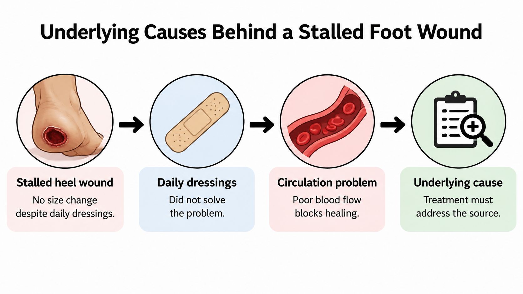
Say a heel wound stops shrinking despite careful dressing changes. The family tries another dressing, but the wound still looks unchanged. If the heel isn't receiving enough blood, neither dressing can correct the main problem.
A useful way to understand this is to think about patching a damaged wall while a pipe continues leaking behind it. Repairing the surface matters, but the repair won't last until someone addresses the source.
The diagram illustrates why looking beyond the dressing matters.
Different causes need different responses
More than one condition can affect the same foot. Someone may have reduced sensation, inadequate circulation and pressure from footwear together, so identifying a single contributor doesn't end the assessment.
| Underlying cause | How it stalls healing | Typical clue |
|---|---|---|
| Diabetic neuropathy | Loss of protective sensation allows repeated injury to go unnoticed | Numb feet, calluses over pressure points |
| Peripheral arterial disease | Reduced blood flow limits oxygen and nutrient delivery | Cold foot, walking-related leg pain; weak pulses on examination |
| Chronic infection or bone involvement | Persistent infection damages tissue and interferes with repair | Drainage or redness that returns after treatment |
| Pressure and repetitive load | Standing, walking or prolonged contact repeatedly injures the wound | Wound beneath the ball of the foot or on the heel |
| Venous disease | Poor return of blood contributes to swelling and skin damage | Leg swelling, varicose veins, brown skin near the ankle |
| Poor glucose control | High blood glucose interferes with immune function and tissue repair | Persistently high glucose readings |
| Malnutrition | Inadequate protein and nutrients limit tissue rebuilding | Unintended weight loss, poor appetite |
These clues guide an examination; they aren't proof of a diagnosis. For example, a wound near the ankle with swelling may involve venous disease, but clinicians still need to check arterial circulation before choosing compression treatment.
Match the questions to your history
If you have diabetes and numbness, make protective sensation and pressure relief priorities. If you smoke, have known vascular disease or experience leg pain while walking, make sure the assessment includes arterial blood flow. Recurrent drainage or a deep wound warrants attention to infection and possible bone involvement.
The range of wound conditions that merit assessment extends beyond diabetes. People with limited mobility can develop heel pressure injuries, and traumatic wounds, surgical wounds and inflammatory conditions such as vasculitis can also stall.
Tell the clinician about reduced appetite, recent weight loss, swelling and difficulty using prescribed footwear. Those details may change the plan as much as the wound's appearance does.
Circulation and Nerve Tests Clinicians Use
Clinicians combine tests because no single result describes the whole foot. A blood-pressure measurement helps assess circulation, a sensation test identifies reduced protection against injury, and imaging addresses a different question, such as whether deeper structures are involved.
A reassuring result on one test doesn't cancel concerning findings elsewhere. For example, stiff, calcified arteries can make ankle pressure difficult to interpret, particularly in people with diabetes. Toe measurements and skin oxygen testing may help clarify the picture.
What each test adds
Exact interpretation belongs with the treating clinician. The wound's location, symptoms, examination and other test results matter alongside the reading.
| Test | What it measures | Reference value or interpretation |
|---|---|---|
| Ankle-brachial index | Ankle blood pressure compared with arm blood pressure | A low result suggests restricted arterial flow; an unusually high result may reflect stiff arteries |
| Toe pressure | Blood pressure measured at a toe | Helps assess whether circulation is adequate for healing, particularly when ankle readings are unreliable |
| Transcutaneous oxygen measurement, or TcPO2 | Oxygen reaching the skin beneath a sensor | Lower readings raise concern about local oxygen delivery |
| Monofilament test | Whether the foot detects a standardized light touch | Missed touches at tested sites suggest reduced protective sensation |
| Probe-to-bone test | Whether a clinician can reach bone through the wound | A positive result raises suspicion of bone infection but doesn't establish it alone |
| Imaging, such as X-ray or MRI | Bone changes, deeper infection or a retained foreign body | Concerning findings guide further investigation and treatment |
The monofilament test isn't the same as asking whether your foot feels numb. You may notice ordinary touch yet lack enough protective sensation to recognize harmful pressure.
Likewise, the probe-to-bone test isn't a home check. A trained clinician performs it and interprets the result alongside the rest of the examination.
Ask for the actual test results, not just “the circulation looks okay.” Keep the report with your wound photographs and treatment notes.
When results don't agree
A foot can look reasonably pink and still have circulation problems. Clinicians may request Doppler studies or further vascular assessment when symptoms, healing progress and initial measurements don't fit together.
Useful questions include: Was the ankle measurement reliable? Was toe pressure assessed? Did the sensation examination show loss of protection? Record the answers so another clinician can understand what has already been checked.
How Clinicians Assess the Wound Step by Step
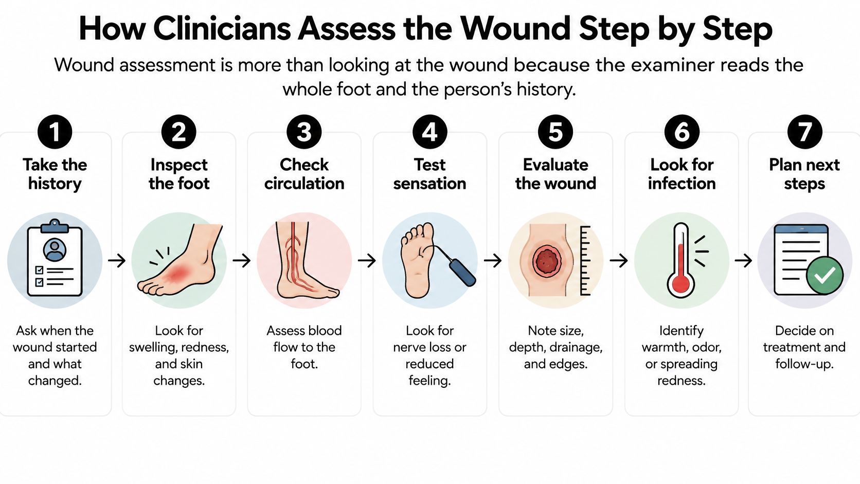
A proper wound assessment includes the whole foot and the person's medical history. The clinician is looking for an explanation that connects the wound's location, tissue appearance and treatment response with circulation, sensation and daily activity.
The assessment follows a structured process, although clinicians may change the order to address an urgent finding first.
From history to a working explanation
- Take the history. The clinician asks when the wound started, what caused it, how it has changed and which treatments have been tried. Glucose control, smoking, previous wounds and medicines that affect bleeding or immune function can alter decisions; a common omission is not mentioning a treatment that was difficult to follow.
- Measure the wound. Length, width and depth are recorded in centimetres, with photographs when appropriate. Consistent technique matters because a different measuring angle can make an unchanged wound appear smaller.
- Describe the tissue. Healthy red granulation tissue, yellow slough and black necrotic tissue suggest different needs. The clinician also looks beneath overhanging edges when safe, because a small opening can hide a larger pocket of tissue damage.
- Look for infection. The assessment includes drainage, redness, warmth, swelling and tenderness, together with general symptoms such as feeling unwell. A common mistake is assuming that the absence of pain or fever rules infection out.
- Assess possible bone involvement. When appropriate, the clinician gently checks whether a sterile probe reaches a hard surface consistent with bone. That finding changes the level of concern, but further assessment is needed rather than treating it as a diagnosis by itself.
- Examine the rest of the foot. Pulses, temperature, colour, sensation, deformities and calluses all provide context. Checking only the open wound can miss a tight shoe, a pressure point or an absent pulse elsewhere on the foot.
- Choose the next investigation. Vascular studies, imaging or tissue sampling are selected to answer the questions raised by the examination. Ordering a test without a clear question can delay the decision that matters.
What meaningful progress looks like
Progress means more than a cleaner-looking photograph. Clinicians look for decreasing wound dimensions, healthy tissue filling the wound bed, advancing skin at the edges and no new surrounding breakdown.
Bring the footwear and pressure-relief device you use, not just a description of them. If you can't safely manage a device at home, say so. A workable treatment plan must account for mobility, balance, dressing support and the help available between visits.
Treatment Choices and When Surgery Is Considered
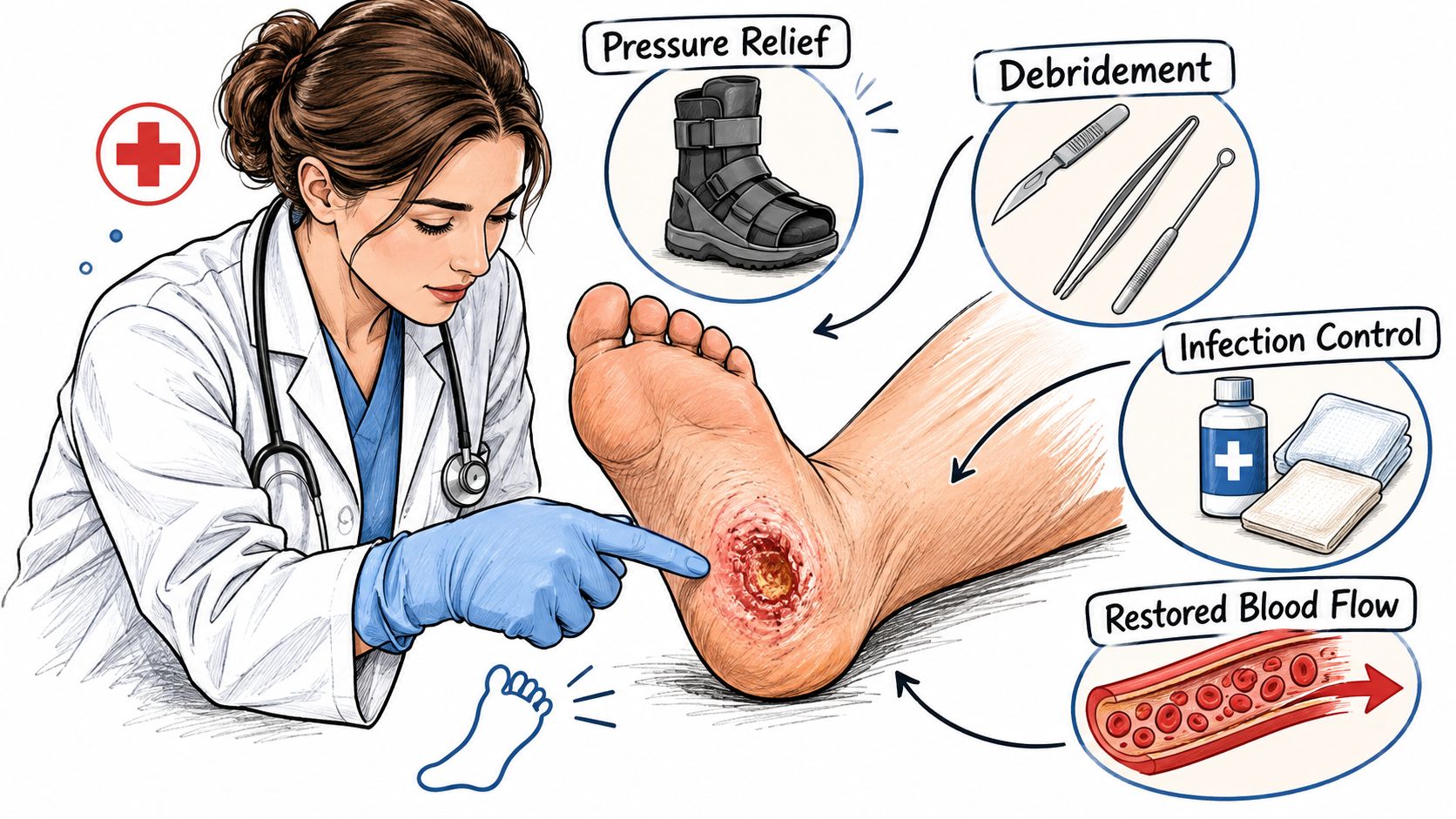
Treatment starts by addressing what prevents repair: pressure, dead tissue, infection and inadequate blood flow. For a wound on the weight-bearing surface of the foot, pressure relief may be more important than changing the dressing brand.
A clinical examination helps determine which treatment should come first.
Pressure relief and local wound care
For an appropriate diabetes-related plantar forefoot or midfoot ulcer, a non-removable knee-high offloading device reduces repeated loading during walking. A removable device may be selected when the non-removable option isn't tolerated or is contraindicated. Device selection must account for infection, circulation, drainage, balance and the need to inspect the wound.
A device that prevents wound access isn't appropriate when frequent examination is necessary because of significant infection, ischemia or heavy drainage. Don't improvise pressure relief by cutting a hole in ordinary footwear or adding padding without guidance; poorly positioned padding can create another pressure point.
Debridement removes selected dead or unhealthy tissue. Its timing and method depend on blood supply and infection, and dry black tissue on a poorly perfused foot shouldn't be cut away at home.
Dressings are then matched to drainage and tissue needs. The aim is a protected, appropriately moist environment without allowing surrounding skin to become waterlogged; more absorbency isn't automatically better for a dry wound.
The following video offers additional context for wound-care discussions.
Infection, blood flow and surgical decisions
Clinicians diagnose infection from the examination, not a positive culture alone. When sampling is needed, tissue from the cleaned, debrided wound base is generally more informative than a superficial swab.
The NICE diabetic foot recommendations support urgent specialist involvement for serious infection; early surgery within 24–48 hours may be indicated alongside antibiotics to remove infected or necrotic tissue. Deep infection, gangrene and arterial disease require coordinated surgical and vascular decisions.
When circulation is inadequate, the vascular team considers whether blood flow can be restored. Urgent drainage and treatment of infection may need to proceed alongside that assessment rather than waiting for every investigation.
Surgery isn't a last resort after dressings fail. It may be necessary for source control, removal of dead tissue or correction of a problem that prevents closure. Conversely, an advanced wound-care assessment may support continued nonsurgical treatment.
Ask: What is the first priority for this wound? Then establish what measurable change should occur and when the team will review the plan.
Getting a Specialist Review and Acting This Week
A wound that has stopped improving deserves a decision about specialist review, rather than another open-ended course of unchanged care. Recurrence, persistent unhealthy tissue or findings that suggest deeper damage strengthen the reason to escalate. Urgent symptoms should go directly to urgent or emergency care, not into a routine referral queue.
Primary care clinicians, podiatrists, vascular surgeons, home health nurses and skilled nursing teams can help coordinate that review. You don't need to arrive with a diagnosis. A clear account of what's changed, what's been tried and what's preventing you from following the plan is useful.
What a specialist review can clarify
At Wound Services, we review the patient's history, wound images, imaging and response to treatment. Our wound surgeon can examine the wound by video, with evaluations usually offered within days; a surgical wound-care nurse then validates the findings in person.
We consider findings such as no measurable size reduction, dead tissue, persistent undermining, recurrent cellulitis, exposed tendon or bone, persistent slough and repeated wound breakdown. Undermining means tissue damage extending beneath the skin beyond the visible opening, something a photograph alone may not reveal.
Our escalation pathway for surgical wound evaluation supports decisions about whether another level of assessment is appropriate. Evaluation doesn't automatically lead to surgery, and there isn't a required waiting period before requesting specialty input.
If intervention is appropriate, the surgeon who evaluates the wound also operates and follows healing afterward. We coordinate recommendations with the existing care team rather than replacing the clinicians already helping the patient.
The purpose of referral is to resolve an unanswered question, not to commit you to a procedure.
A practical plan for this week
- Create a baseline. Photograph the wound and record its visible dimensions, drainage and surrounding skin changes. If you can't see the sole or heel safely, ask a caregiver to help rather than twisting or balancing in a way that risks a fall.
- Respond to urgent changes immediately. Use same-day assessment for concerning local changes and emergency care for systemic illness or sudden circulation changes. Don't postpone care while collecting photographs, records or insurance information.
- Arrange a focused visit. Ask the current clinician to review circulation, protective sensation and the pressure-relief plan. Bring the device you use and explain any difficulty with walking, transfers or wearing it consistently.
- Request specialist input when progress is absent. Bring evidence of the stalled trend and ask which unresolved issue needs further assessment. You can compare the proposed options, including continued conservative care, additional testing and procedural treatment.
- Prepare a portable wound record. Include the medication list, allergies, recent test results, imaging reports and previous treatment instructions. Write down who will make the next decision and how you'll receive the result.
Before leaving an appointment, confirm the follow-up plan and whom to contact if the wound changes. A plan without a review point can leave patients repeating care without knowing whether it's helping.
You don't have to wait for a wound to become larger, painful or infected before asking for an evaluation. Asking while you're uncertain is a reasonable part of protecting your foot.
---
We offer virtual and in-person specialty evaluation for complex, stalled and recurrent foot wounds, with coordinated surgical care when clinically appropriate. Visit Comprehensive Wound Services to request an evaluation and help clarify whether your wound needs further testing, a revised treatment plan or surgical assessment.