
Purulent drainage is thick, opaque fluid, usually yellow, green, or tan, made up of pus, and it is a warning sign that needs clinical assessment rather than a diagnosis on its own. Surgical-site infections occur in 1%–3% of people undergoing surgery, so this fluid is uncommon enough to take seriously and common enough that a caregiver should know how to read it.
Is the yellow stain on the dressing a sign that the wound is infected, or is it one clue that only makes sense alongside depth, timing, and how the wound looked yesterday? A lot of advice treats cloudy drainage as an automatic infection label. That shortcut skips the essential question. Pus indicates a fight is underway. It doesn't tell you how deep the fight is, whether fluid is trapped in a pocket, or whether another antibiotic will change anything.
## Table of Contents - Purulent Drainage Defined in Plain Terms - Telling Purulent Drainage Apart from Normal Wound Fluid - The colour trap - What Purulent Drainage Can Indicate - Warning Signs That Need Urgent Care - Seek urgent or emergency care - Contact the clinician the same day - How to Document Drainage Between Visits - Why Drainage Persists Despite Antibiotics - What a stalled course usually means - Confirm the Next Step With a Wound Clinician
Purulent Drainage Defined in Plain Terms
Purulent drainage from wound care is thick, opaque fluid, usually yellow, green, or tan, and it often has an odor. It is made of white blood cells, bacteria, and tissue debris. Picture pus as leftover debris from the wound's cleanup crew after a fight with germs. The colour tells you a fight is happening. It doesn't tell you how large that fight is, or whether it is winning.
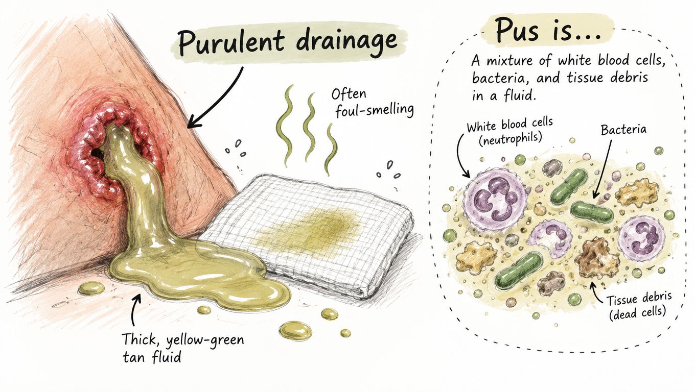
The Cleveland Clinic reported in 2025 that the U.S. Centers for Disease Control and Prevention estimates surgical-site infections occur in 1%–3% of people undergoing surgery, and it lists purulent drainage as a symptom of surgical-wound infection. Most surgical wounds never produce pus. A pad that looks off still needs a person to look at the whole wound, not a label based on colour alone.
You'll meet this fluid on incisions after surgery, on traumatic cuts, in pressure injuries, and inside abscesses. In every one of those places it is a warning sign, not a finished diagnosis. A small stitch line and a deep pressure injury can both stain gauze yellow. They don't carry the same risk.
Clinicians read three things together.
- What the fluid looks like: thickness, colour, and smell.
- When it appeared: day 2 isn't day 12.
- What the surrounding skin is doing: warmth, redness, tenderness, or edges pulling apart.
Pus is one clue among several. The timing and the skin around the wound decide whether you keep watching or you call.
Telling Purulent Drainage Apart from Normal Wound Fluid
Drainage has four common looks, and only one of them is pus.
| Drainage Type | Appearance | Usually Expected? | Concern Signal |
|---|---|---|---|
| Serous | Clear, thin, watery | Expected in small amounts during early healing | Volume rising or fluid turning cloudy |
| Serosanguineous | Pale pink to straw-colored, thin | Common in the first postoperative days | Turning cloudy, thick or foul-smelling |
| Sanguineous | Bright red, thin | Expected shortly after injury or surgery | Ongoing heavy bleeding or clots that keep forming |
| Purulent | Thick, opaque, yellow, green or tan; may smell foul | Not expected as routine seepage | Any new purulence needs prompt clinical assessment, especially with fever, spreading redness or pain |
Serous fluid is the thin, watery plasma of early healing. Serosanguineous fluid is that same fluid with a little blood, so it looks pale pink or straw-colored in the first days after an operation. Sanguineous fluid is bright red and thin, the fresh blood you expect right after injury or surgery, and it should ease rather than keep soaking pads. Purulent fluid is the thick, opaque one. It isn't routine seepage.
The colour trap
Yellow material in a healing wound can be fibrin or slough, not pus. Cloudy fluid isn't automatically infected. Colour alone can't settle the question, because the same shade can mean different things on day 2 and on day 12.
A 2025 Canadian best-practice resource indicates that minimal-to-moderate sanguineous or serous exudate can occur during the first several postoperative days, whereas serous or purulent exudate beyond day 9 warrants infection assessment. Reading also depends on quantity, change from the wound's own baseline, odor, pain, warmth, redness, wound separation, and how the person feels overall.
Say a hip incision weeps pale pink fluid on day 2, then on day 11 the pad is thicker, yellow, and smells different from yesterday. That change matters more than any single glance. Chronic wounds make this harder still, because bacteria often live on the surface without invading tissue. Drainage there doesn't, by itself, prove invasive infection.
Practical rule: Compare today's drainage with your last record. A change from that wound's baseline beats any one reading.
Unnecessary antibiotics can cause side effects and feed resistance. Treating thick fluid as harmless can delay care. Infection is a clinical call, and systemic antibiotics are generally reserved for infection that has become systemic. Local care can be enough when the problem stays local. Write down colour, consistency, odor, and volume, look at the skin, and call promptly if drainage is increasing or arrives with fever, spreading redness, worsening pain, warmth, dead tissue, undermining, or edges that open.
What Purulent Drainage Can Indicate
Purulent fluid can point to infection in the skin, in deeper tissue, or in a collected pocket. It doesn't set the severity by itself.
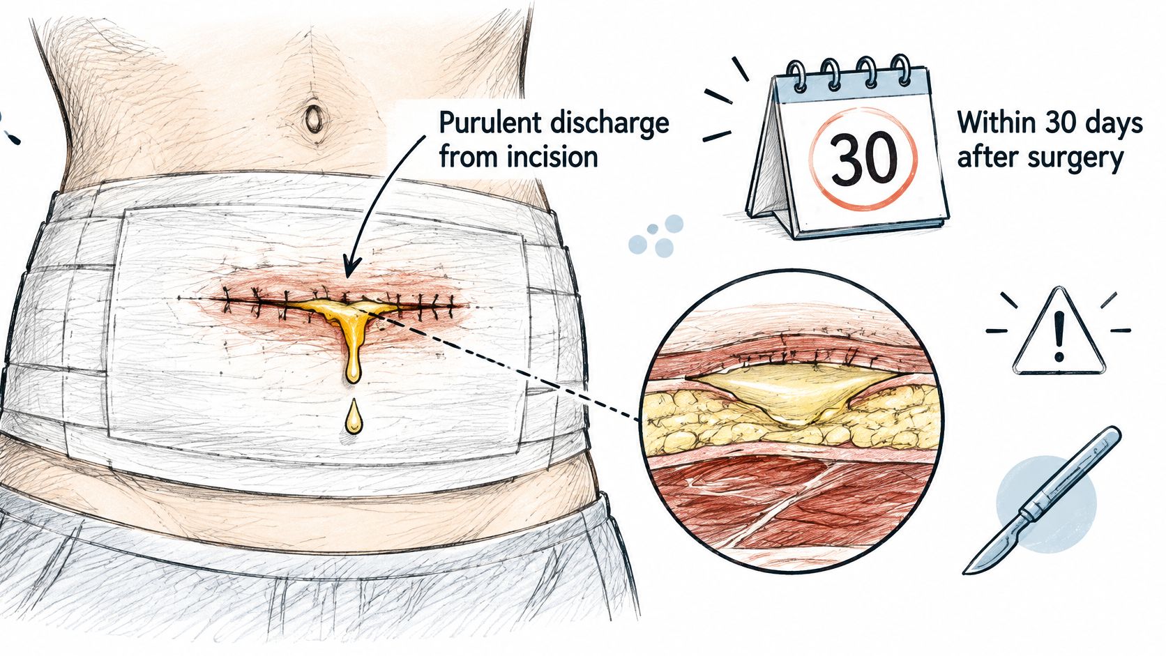
CDC surveillance definitions classify a superficial incisional surgical-site infection when purulent discharge comes from the incision within 30 days after surgery, even without laboratory confirmation. Deep incisional infection is similarly supported by purulent drainage from the deeper portion of the incision. Other qualifying findings include fever above 38°C, localized pain or tenderness, wound separation, or an abscess found during examination, reoperation, imaging, or pathology. The CDC estimated 110,800 inpatient-surgery-associated surgical-site infections in the United States in 2015. That surveillance window classifies infections for tracking. It isn't a cutoff that says infection can't exist outside it, and the earlier day-9 cue is a reason to assess a wound, not a rule that infection starts on a calendar date.
| Location or Pattern | What It May Indicate | Typical Next Step |
|---|---|---|
| Skin-level incision, drainage within 30 days of surgery | Superficial incisional infection | Clinical wound exam, local care, timely review |
| Deeper incision with tenderness or wound separation | Deep incisional infection involving fascia or muscle | Examination, imaging and possible drainage |
| Firm, tender pocket or swelling that fluctuates | Undrained abscess | Incision and drainage, often with culture |
| Persistent drainage with dead space or retained material | Undrained collection or retained material | Source-control assessment |
| Long-standing chronic wound with ongoing drainage | Colonization or chronic infection | Reassessment by a wound clinician |
Say someone is six days after a hernia repair. The drainage turns thicker and yellow, the skin beside the scar is warm, and a tender pocket sits under the dressing. That pattern points toward a collected or deeper problem, not ordinary skin-level seepage. Pus on the pad doesn't tell you which row of the table you're in. Depth, timing, and the way the wound feels do that work.
For pressure injuries, diabetic ulcers, and traumatic wounds, incision criteria don't apply. Read depth, trend, and the surrounding skin against the same patterns, and expect a wound clinician to judge surface colonization against invasive infection.
Warning Signs That Need Urgent Care
No single sign proves infection. Groups of signs tell you how fast to move.
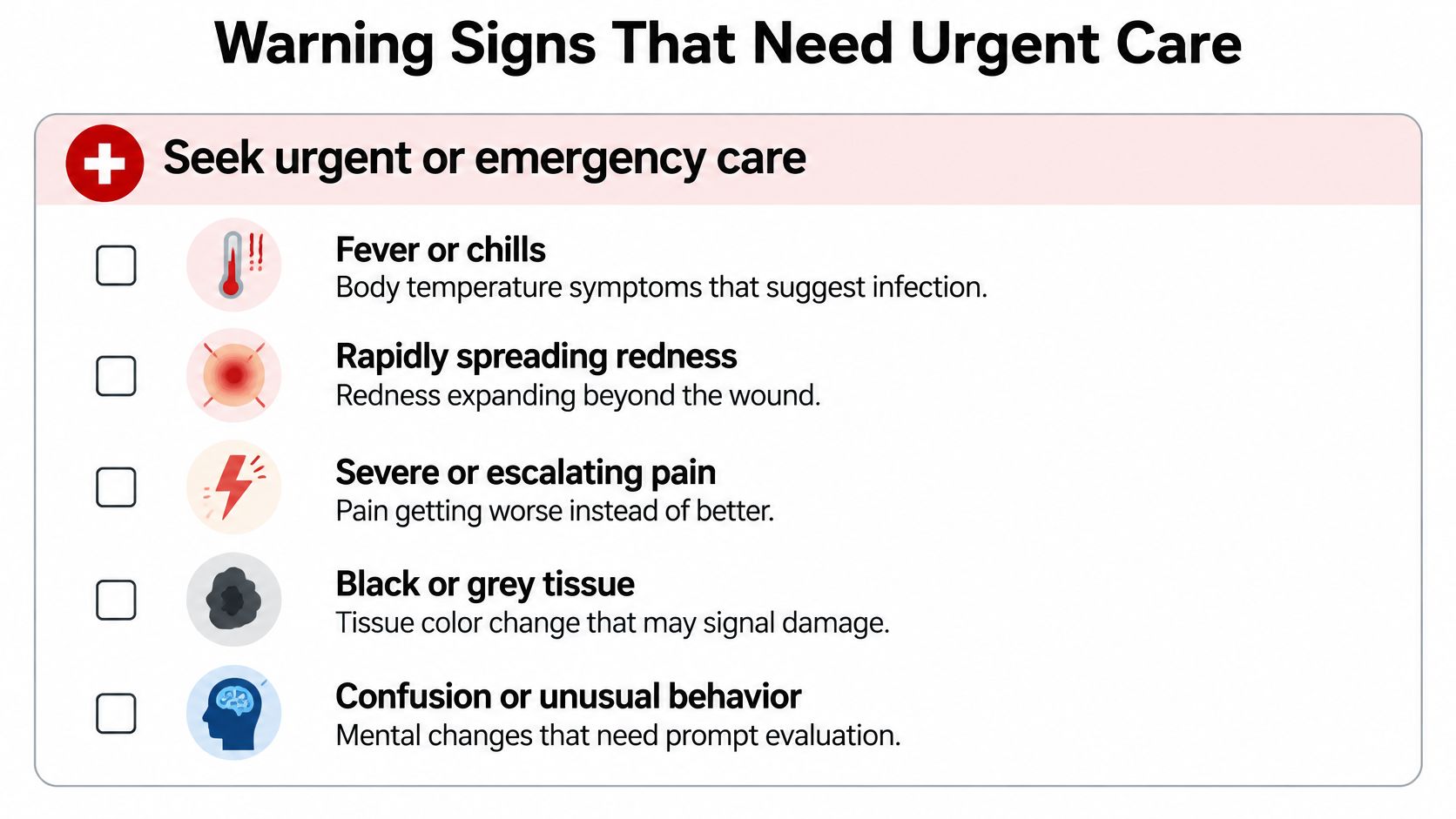
Seek urgent or emergency care
- Fever or chills: The body is reacting beyond the wound.
- Rapidly spreading redness: Redness is moving outward, not staying at the edge.
- Severe or escalating pain: Pain is climbing instead of settling.
- Black or grey tissue: Colour like this can mean tissue is dying.
- Confusion, unusual drowsiness, or new weakness: Thinking or strength has changed.
- Exposed bone or tendon: Deeper structures are open to the air.
- Can't keep fluids down: Hydration is failing along with the wound.
Contact the clinician the same day
- Drainage increasing from one check to the next: Volume or thickness is worse than your last note.
- New or worsening odor: The smell changed, not just the colour.
- Edges opening: The wound is separating.
- Pain that changes character: A new kind of pain, even if it isn't severe yet.
- Purulence that returns after treatment: The fluid came back while care was already underway.
If you've had surgery, the team that operated is the first call for questions about that incision. If you can't tell which group fits, treat the question as urgent and call. Our escalation pathway is built for wounds that need a faster clinical look than another day of watching.
When you're unsure which group applies, don't wait overnight to decide. Call.
How to Document Drainage Between Visits
A short log turns a vague worry into a trend a clinician can use. It doesn't replace an exam.
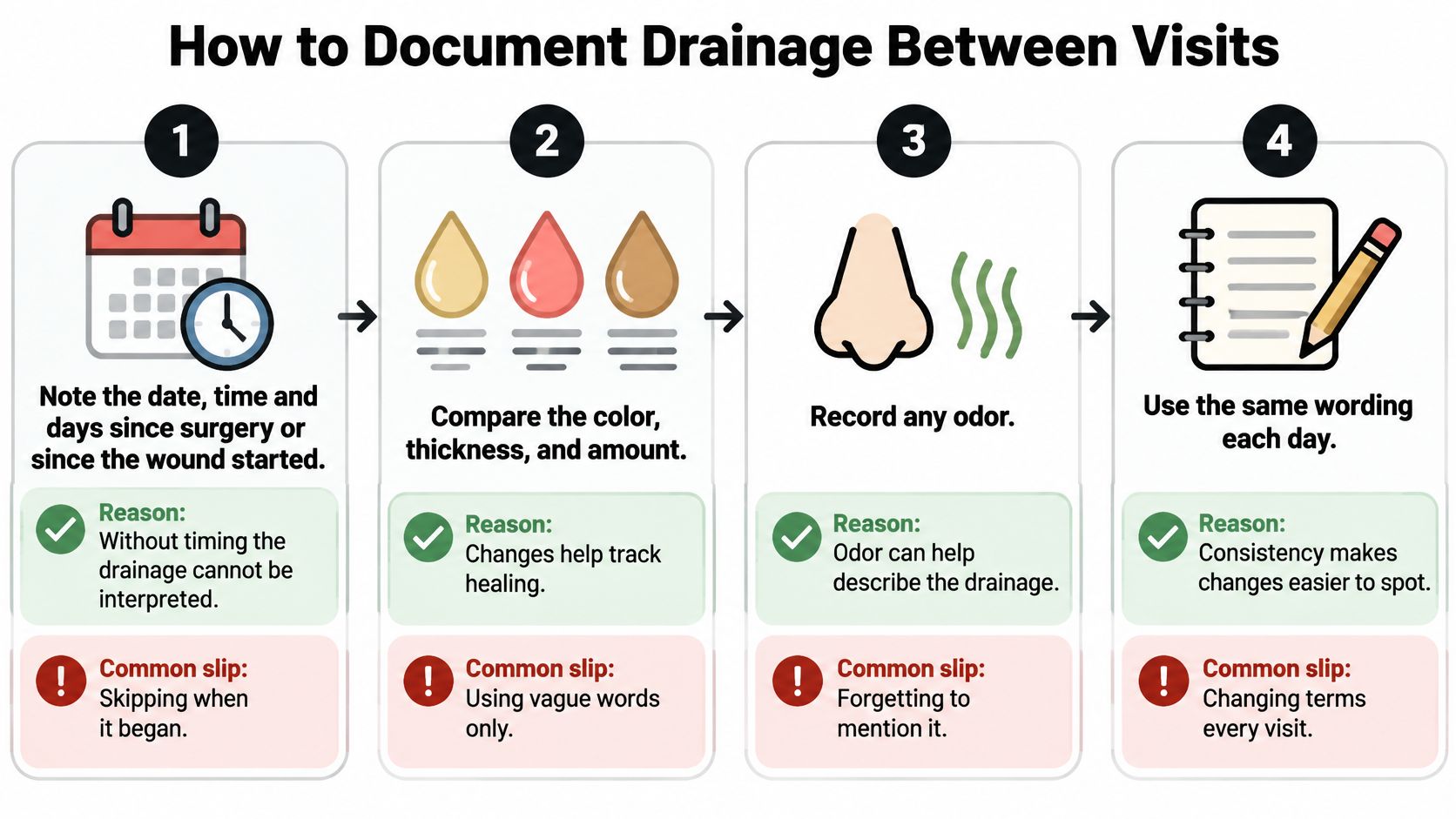
- Note the date, time, and days since surgery or since the wound started. Without timing, nobody can interpret the fluid. The common slip is writing "some drainage" and skipping when it began.
- Compare volume and colour with your last record. A change from that wound's baseline matters more than one glance. Don't rely on memory from yesterday's dressing change.
- Describe colour, consistency, and odor in plain words. "Thin and clear," "thick and creamy," or "sweet-smelling" is more useful than guessing the diagnosis. Skip words like "infected" unless a clinician has already said that.
- Inspect the skin for spreading redness, warmth, swelling, or red streaks. If redness is present, mark the outer edge with a skin-safe pen and note whether it moved. Don't mark over broken skin.
- Rate pain and note whether it is increasing or new. A jump from mild ache to throbbing at rest is a change, even if the pad looks the same.
- Take your temperature and write the number down. One reading is a data point. A rising pattern is a reason to call.
- Look for opening, black or grey tissue, or a firm pocket. Don't press hard. Squeezing the wound to "see more drainage" can push material deeper and irritate tissue.
- Decide. If drainage is increasing or any warning sign is present, call the clinician the same day.
Between visits, photos in the same light help, and remote wound monitoring works best when the notes use the same words each day. The log is for the clinician. It isn't a substitute for assessment, and it isn't a reason to wait once a warning sign shows up.
Why Drainage Persists Despite Antibiotics
Why might pus return even while you are taking antibiotics? Drainage is a clue, not a diagnosis. Its depth, timing, and trend help explain what may be happening, and more antibiotics may miss the source.
Persistent drainage can be associated with an undrained pocket, dead space, foreign material, dead tissue, a wound that has opened, resistant organisms, or infection deeper than the skin. An exam may check for fluid pockets, tissue damage, poor blood flow, or undermining. A clinician may also consider a sample for culture or imaging to help guide treatment. Antibiotics may not resolve an infection if a pocket of pus remains undrained.
Antibiotics are like a tool that cannot reach behind a closed door. If pus is trapped in a pocket, a clinician may need to drain it; the right treatment depends on the wound and the assessment. The Infectious Diseases Society of America's guidance on skin and soft-tissue infections describes drainage as the main treatment for many abscesses, carbuncles, and large furuncles. Recurrent abscesses may need drainage and culture.
What a stalled course usually means
If drainage is not improving, contact your clinician for reassessment rather than extending or repeating antibiotics on your own. Do not change your dose, use leftover medication, or start another course without medical advice. A clinician may need to reconsider the wound's depth, whether material is trapped, and whether a culture would help identify the organism. Molecular testing for wound infections can help identify microbial DNA and selected resistance markers as part of that assessment. Testing does not drain a pocket that needs source control.
Worsening or returning drainage while you are taking medication is a reason to call. Seek same-day care for fever, chills, rapidly spreading redness, severe or increasing pain, black or grey tissue, confusion, weakness, exposed bone or tendon, or trouble keeping fluids down. Don't wait until the medication is finished if these signs appear.
Confirm the Next Step With a Wound Clinician
Bring a drainage log with dates, photos taken in the same light, your current medication list, every antibiotic with start and stop dates, and any culture results you already have.
Ask three questions. Is this drainage superficial or deeper? Does it need culture or imaging? Does it need drainage or other source control?
Call the surgeon who operated for incision questions. Call your primary clinician for general concerns. Use urgent care or emergency care for any sign in the urgent group. For complex, stalled, or recurrent wounds, a wound specialist is one option, not a required step. We review history, images, and the wound by video, usually within days, and the surgeon who evaluates the wound is the surgeon who operates when intervention is appropriate, and who follows the healing after.
Surgical evaluation is worth discussing when a wound shows no measurable size reduction, necrotic or dead tissue, persistent undermining, infected tissue or recurrent cellulitis, exposed tendon or bone, persistent slough, recurrent breakdown, or minimal improvement despite appropriate evidence-based care. Book the visit with the log in hand, and treat any urgent sign as a reason to call the same day.
---
Wound Services evaluates complex, stalled, and recurrent wounds, including purulent drainage that may need source control, debridement, or incision and drainage when that is the right next step. Virtual and in-person review can sort a surface problem from a deeper one before anyone assumes another antibiotic is the answer. Visit Comprehensive Wound Services to arrange that review.