
You're on day 18 after surgery, the dressing comes off, and the incision looks almost the same size it did last week. Drainage is still there. Pain hasn't clearly eased. You're trying to decide whether to wait for the routine visit or call someone today. A surgical wound not healing is a pattern, not one bad glance. You judge it by whether the wound is getting smaller, staying closed, and calming down. The response depends on which sign you see and how fast it showed up.
## Table of Contents - How Long a Surgical Wound Should Take to Heal - What each phase should look like - Warning Signs and How Urgent Each One Is - How signs stack - Why Surgical Wounds Stall in the First Place - Four families of causes - Infection, Biofilm and the Limits of Antibiotics - What points to infection - What Specialist Treatment Changes That Dressings Cannot - Other decisions a specialist may make - What to Do at Home While the Wound Is Being Watched - Protect the seam from strain - Virtual Review Versus In-Person Wound Assessment - What to send if you can't easily travel - Decide Whether to Book a Specialty Evaluation Now - What to have ready
How Long a Surgical Wound Should Take to Heal
On day 18, someone looks at an abdominal incision and can't tell it apart from the photo taken a week earlier. The edges are pink. A little fluid is still on the gauze. That stall is the question, and the answer comes first. A surgical wound should show measurable progress within about two to four weeks. A wound that hasn't begun to improve by then is nonhealing and warrants specialty review. The Wounds UK meeting report describes concern when healing stalls or regresses, and it treats a wound that hasn't begun to improve after roughly 2–4 weeks as nonhealing.
What each phase should look like
| Healing phase | Typical timing | What progress looks like |
|---|---|---|
| Inflammation | Days 0 to 4 | Redness and swelling settle; drainage is clear and decreasing |
| Proliferation | About day 4 to weeks 3 to 4 | Pink granulation tissue fills the base; edges begin to close; size shrinks each week |
| Maturation | Weeks to 12 months or more | Scar strengthens, flattens and fades in color |
| Stall threshold | About 2 to 4 weeks | No measurable improvement yet: request specialty review |
Inflammation is the noisy first stretch. Redness and swelling should settle, and drainage should turn clear, then lessen. Proliferation is the working stretch. Pink granulation tissue fills the base, the edges start to move inward, and the wound should be smaller from one week to the next. Maturation is slow. The scar strengthens, flattens, and fades over months, sometimes past a year. You won't see week-to-week shrinkage then the way you should in the first month.
Stalling is a trend, not one rough morning. A wound that stops contracting, reopens more than once, or looks larger despite appropriate care matters more than its size on a single day. Speed also changes with location, tension across the incision, diabetes, smoking, and how much protein the person is eating.
Practical rule: Measure length, width, and depth on the same day each week, with the same ruler and the same lighting. A shrinking set of numbers is progress. A flat set after two to four weeks is the signal to ask for specialty review.
Warning Signs and How Urgent Each One Is
Match the sign to a clock. Some changes need a call today. Others can be tracked at home if nothing else is worsening.
| Symptom | What it may signal | Suggested response timing |
|---|---|---|
| Fever, chills or feeling generally unwell | Systemic infection | Urgent evaluation the same day |
| Redness, warmth or swelling spreading past the edge | Spreading infection or cellulitis | Same-day clinician call |
| Pus, cloudy or foul-smelling drainage | Surgical-site infection or abscess | Same-day clinician call |
| Edges separating or the wound reopening | Dehiscence | Same-day clinician call |
| Exposed tendon, bone or deeper tissue | Deep compromise | Urgent evaluation the same day |
| Black or dark dead tissue (eschar) | Necrosis | Prompt specialty review |
| Undermining or tunnels felt beneath the edges | Dead space or hidden collection | Prompt specialty review |
| Pain that grows rather than eases after the first days | Infection or poor blood supply | Same-day clinician call |
| No measurable size reduction after 2 to 4 weeks | Stalled healing | Specialty evaluation |
| Dry, unchanged dressing with normal healing signs | Usually normal healing | Monitor and track measurements |
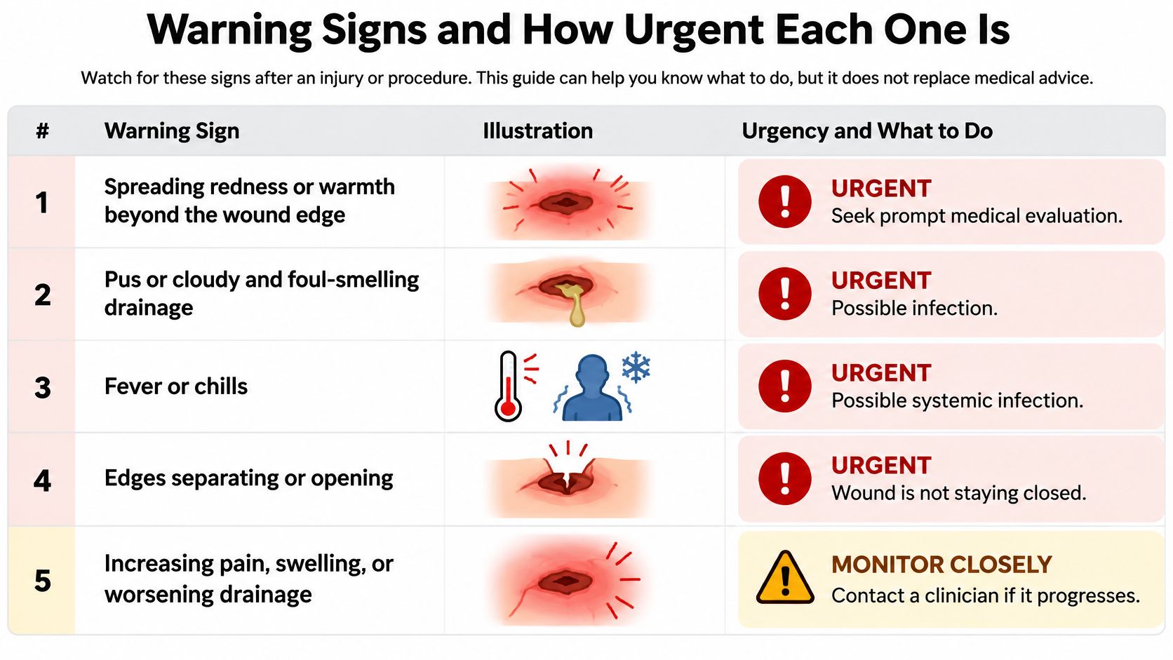
The table supports judgment. It doesn't replace it. A single lower-urgency finding can become urgent the moment it shows up with another one. Flat measurements plus a new fever are not a "watch another week" problem. Black tissue plus a new tunnel under the edge is not a dressing problem.
How signs stack
We use eight indicators when a wound may deserve surgical evaluation: no measurable size reduction, necrotic or dead tissue, persistent undermining, infected tissue or recurrent cellulitis, exposed tendon or bone, persistent slough, recurrent wound breakdown, and minimal improvement despite appropriate evidence-based care. One of those, clearly present, is enough to book a review. Two together should move the call to the same day.
Say the wound is slowly smaller, cool, and the person has no fever. Keep the weekly measurements and the planned dressing changes. Now give that same wound a fever and redness that has marched past the ink line you drew yesterday. That pair needs a clinician the same day, even if the wound was shrinking on Monday.
Pain that grows after the first few days is easy to explain away as "healing hurts." Early soreness should ease. Pain that climbs, especially with swelling or heavier drainage, belongs on the same-day row. A dry dressing, edges that stay together, and redness that is fading can stay on the monitor row.
A normal-looking dressing doesn't cancel a fever, a new opening, or tissue you can see that shouldn't be visible.
Why Surgical Wounds Stall in the First Place
A closed incision is a repaired seam. It holds only when the edges stay aligned, the suture isn't asked to carry too much load, and the tissue underneath still gets blood.
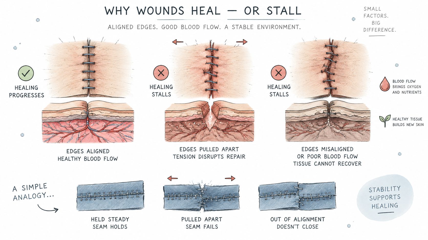
When a seam fails, clinicians call the partial or complete separation of previously closed layers surgical wound dehiscence. A systematic review and meta-analysis of abdominal surgery pooled 741,118 patients and found a worldwide 30-day cumulative incidence of about 1% after laparoscopic or open abdominal surgery (95% confidence interval 1%–1%). Hepatobiliary surgery had the highest reported incidence in that review, about 3% (95% confidence interval 0%–8%). The authors cautioned that the estimate may understate the true burden, because most studies were retrospective and definitions weren't consistent. That 30-day figure is a population benchmark, not a forecast for one person.
Four families of causes
Local problems sit in the wound itself. Infection, poor blood supply, a pocket of dead space, retained suture or mesh, leftover contamination, and too much tension can each stop the edges from knitting.
Systemic problems change the person's ability to build tissue. Diabetes, obesity, malnutrition, smoking, immunosuppression, and corticosteroid use all slow repair, even when the operation was clean.
Procedural problems raise the starting risk. Emergency surgery, a heavily contaminated field, and a long operation leave more injured tissue and more bacteria than a short elective case.
Mechanical problems arrive later, at home. Coughing, vomiting, heavy lifting, and abdominal pressure pull on a seam that isn't strong yet.
Picture an incision that looks sealed at the top and wet at the bottom. After a hard coughing spell, the lower end gaps a few millimeters and the drainage increases. Strain opened a weak spot. Fluid and bacteria then keep that spot from sticking. Fixing only the cough, or only the dressing, misses half the problem.
A short explanation of how a closed wound can separate under strain helps caregivers see why "it was fine this morning" can change by evening.
One cause rarely acts alone. Naming the dominant one decides the next move. Strain needs support and activity limits. Dead tissue needs removal. A deep pocket needs drainage. A person who is malnourished needs protein and calories while those local problems are treated.
Infection, Biofilm and the Limits of Antibiotics
Antibiotics are not the default fix for a quiet, stalled incision. International postoperative guidance cautions against antimicrobial drugs when there is no active infection. Drugs matter when infection is present. Removing the source often matters as much as the prescription.
Surgical-site infection means bacteria damage tissue, drive drainage and inflammation, pull edges apart, and sometimes track into deeper layers. It is common enough to check every time a wound stops moving. On the World Health Organization surgical-site infection page, a worldwide meta-analysis of adult general-surgery patients put pooled 30-day incidence at 11% (95% confidence interval 10%–13%) across 488,594 patients. A Cleveland Clinic patient page, citing the U.S. Centers for Disease Control and Prevention, places infections at about 1%–3% of people who have surgery. The gap reflects different populations and definitions. Neither number is your personal risk. Both are reasons to look, not to guess.
What points to infection
- Drainage: Pus, cloudy fluid, or a foul smell is not the clear fluid of the first day or two.
- The rim: Warmth, swelling, or redness that spreads past the edge suggests cellulitis.
- The whole person: Fever, chills, or a new sense of being unwell can mean the infection isn't staying local.
- The structure: An abscess, or an incision that opens on its own, means the closure has already lost.
Bacteria can also sit in a biofilm, a sticky film that blunts both antibiotics and immune cells. The surface can look fairly calm while the base never fills in. That is why a leftover antibiotic from an old prescription is a poor plan, and why stopping a prescribed course early is a poor plan too. Spreading cellulitis and systemic signs need antimicrobials and a same-day exam. A collection of pus needs to be opened. Dead tissue needs to come out. Culture or molecular testing is most useful on tissue taken from the right depth, not on a casual swab of the wet surface.
Say the edge is wet with pus but the base is pink and the person has no fever. The plan is local source control and close follow-up. If that same wound has fever and redness climbing the abdomen, the plan adds urgent assessment for deeper spread. Location changes the treatment. A surface problem and a deep one should not get the same response.
What Specialist Treatment Changes That Dressings Cannot
A wound that stops shrinking needs a check for biofilm, dead tissue, foreign material, undermining, and infection. Another brand of dressing doesn't answer those questions.
Sharp or surgical debridement is the step dressings can't copy. It clears necrotic tissue and breaks up the matrix that shelters bacteria, which briefly makes those bacteria easier to treat. Because the film can rebuild quickly, the next steps have to follow at once.
- Remove nonviable tissue with sharp or surgical debridement, which opens a short window when remaining bacteria are more susceptible.
- Clean the wound bed and apply an appropriate topical antimicrobial or antibiofilm agent immediately, before the film re-forms.
- Reassess length, width, depth, tissue quality, exudate, pain, and redness around the wound.
- Repeat the cycle when slough, necrosis, or stalling returns, rather than waiting for a calendar date.
The biofilm and debridement review in PMC is clear that antimicrobial dressings alone shouldn't stand in for source control. If infection spreads beyond the wound, or fever and chills appear, antimicrobials and urgent assessment are required. Tissue samples are most informative after debridement. A swab taken before dead tissue is removed can miss the organisms living underneath.
Other decisions a specialist may make
A hidden fluid pocket may need drainage. Retained suture, mesh, or other material may need removal if it keeps the wound contaminated. Undermining and dead space may need packing, a drain, or closure of the pocket so fluid can't pool. When contamination is heavy, the choice may be delayed primary closure or healing by secondary intention, meaning the wound fills in open rather than being sewn shut again. High-quality evidence still doesn't settle the best moment to start or stop negative-pressure wound therapy, so that device is a tool with a plan, not a guarantee.
Conservative care stays the foundation. There is no required waiting period before an evaluation, and an evaluation doesn't automatically lead to surgery.
Advanced wound care at a visit should include a fresh measurement, a written plan, and a clear checkpoint. You should leave knowing what "better" looks like at the next look, and which change means you shouldn't wait for that date.
What to Do at Home While the Wound Is Being Watched
Home care is the baseline, not a substitute for the urgent signs. Keep the dressing clean and dry, and change it on the schedule you were given. Wash your hands before you touch the incision. Skip creams, hydrogen peroxide, and home remedies nobody prescribed. Those products can irritate tissue that is trying to close.
Protect the seam from strain
- Support the abdomen or the incision with a hand or pillow when you cough, sneeze, or stand up.
- Skip heavy lifting until a clinician clears it. A wound can feel fine and still be weak.
- Ask about vomiting or constipation. Both raise pressure inside the abdomen and tug on the closure.
You can also change the conditions repair depends on. Cut down or stop smoking, because nicotine narrows the blood supply the edge needs. If you have diabetes, keep glucose in the range your clinician set. Eat regular protein and drink enough fluid so the body has material to build tissue. Don't stop a prescribed antibiotic early, and don't start a leftover one because the wound "looks infected."
Each day, note drainage color and amount, pain compared with yesterday, and whether you felt feverish. Once a week, record length, width, and depth. Bring that log to every visit so the trend is visible in minutes. Use these home observations as the trigger to change the plan: fever or chills, redness or warmth past the edge, pus or foul drainage, edges that separate, black tissue, a tunnel under the rim, pain that is rising, or measurements that haven't moved. A dry dressing with fading redness and a smaller wound can stay on the monitoring track.
Virtual Review Versus In-Person Wound Assessment
A virtual review can speed triage, history review, image assessment, and a look at how treatment is working. It can't replace an in-person exam when deep infection, necrosis, dehiscence, exposed bone or tendon, or a debridement decision is in question.
| Assessment task | Virtual review | In-person assessment |
|---|---|---|
| Review history, medications and treatment response | Yes | Yes |
| Assess photographs, measurements and imaging | Yes, if image quality is adequate | Yes, with direct measurement |
| Confirm necrosis, undermining or exposed structures | Limited | Yes |
| Judge deep infection or fluid collection | Limited | Yes |
| Plan debridement or a surgical procedure | Planning only | Yes, with the procedure decided after examination |
| Timing of the review | Usually within days | Scheduled, or sooner when escalation is needed |
A 2025 systematic review in JMIR mHealth and uHealth covered 22 randomized trials and 2,397 participants. Telemedicine improved wound-healing scores, with a standardized mean difference of −1.46, and was linked to lower amputation risk (RR 0.52). The authors stressed substantial heterogeneity and said the evidence isn't strong enough for firm conclusions on definitive healing rates. Virtual care can help. It isn't proof that every wound can be managed on a screen.
We review the history, the wound, imaging, and the treatment response by video, usually within days. A surgical wound care nurse then validates the wound in person when that check is needed, so what was seen on camera is confirmed on the body.
What to send if you can't easily travel
- Photos: Daylight shots with a ruler in the frame, dated, from straight above and from the side.
- Numbers: Length, width, and depth from the same weekday each week.
- The record: A medication list, plus notes on drainage, pain, and any fever.
Fever, pus, exposed tendon or bone, edges that have opened, or a tunnel you can feel are in-person triggers. Nursing-home residents and people without a ride still need those findings seen directly. Video is a fast first step. It isn't a substitute for hands on the wound.
Decide Whether to Book a Specialty Evaluation Now
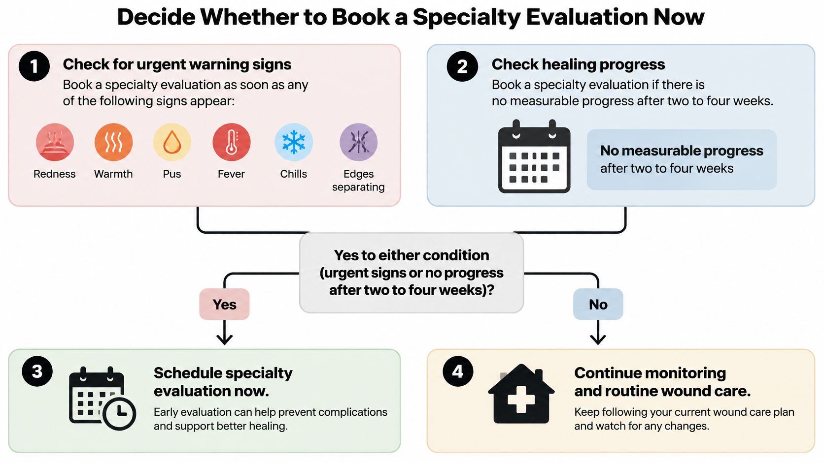
Book a specialty evaluation as soon as any same-day sign appears, or when measurements show no progress after two to four weeks. Waiting for an arbitrary extra month doesn't make a stalled wound safer.
What to have ready
- Write the surgery date and the date you first noticed the wound stop changing.
- Photograph the wound in daylight with a ruler beside it.
- Measure length, width, and depth on the same weekday each week.
- List diabetes, smoking, eating problems, and current medicines, including steroids.
- Record drainage type, a pain score, and any fever.
- Bring the operative note and the antibiotic history if you have them.
A surgical wound evaluation is not an automatic trip to the operating room. The surgeon who evaluates the wound is the surgeon who operates if intervention is appropriate, and who follows healing afterward. If surgery isn't the right step, you leave with a plan and your existing clinicians stay in charge. After an operation, follow-up and recommendations return to that team, and virtual follow-up tracks the wound toward closure. When surgery is needed, a nurse validates the wound in person, transportation is arranged, and you go to our ambulatory surgical center, with same-day discharge home when that is clinically suitable.
Contact your current surgeon or primary clinician this week if the wound is flat. Go to same-day care for fever, spreading redness, pus, a reopening incision, exposed deeper tissue, or pain that is getting worse. Those signs don't wait for a scheduled slot.
---
Wound Services evaluates complex and stalled surgical wounds with virtual review and in-person checks, and we operate only when that is the right next step. If your incision has stopped closing, reopened, or developed a sign that needs a same-day look, you can start with a specialty review instead of waiting through another unchanged week. Visit Comprehensive Wound Services to request an evaluation and bring your photos, measurements, and medication list.