Biofilm was found in 78.2% of chronic wounds examined in a 2017 systematic review and meta-analysis. That finding reframes a wound that won't heal: microorganisms may be living in organized, protected communities even when the surface doesn't look obviously infected.
For patients and caregivers, the important distinction is between controlling biofilm and proving that a treatment helps a wound close. Understanding both makes it easier to interpret test results, follow a treatment plan, and recognize when a stalled wound needs another level of assessment.
## Table of Contents - How Common Biofilm Is in Chronic Wounds - Understanding the detection range - How Biofilm Keeps Chronic Wounds From Healing - Protection changes the response to treatment - Inflammation interferes with rebuilding tissue - How to Spot Biofilm When a Wound Looks Clean - Separate microbial presence from infection - Compare progress, not just appearance - How to Assess a Stalled Wound Step by Step - A repeatable clinical routine - What caregivers can record between visits - How Biofilm Is Treated With Debridement and Topical Care - What each treatment component adds - Match treatment intensity to response - Measure the wound's response - What Biofilm Treatments Can and Cannot Promise - Microbial improvement isn't the same as closure - Promising options still need clinical proof - Decide When a Stalled Wound Needs Specialist Care - Findings that deserve a closer assessment - What specialty review involves - Keep appropriate care going while arranging review
How Common Biofilm Is in Chronic Wounds
Biofilm is a community of microorganisms embedded in a self-made protective matrix that clings to the wound surface. It isn't loose bacteria sitting on top of the skin, and it isn't something caregivers can reliably identify by looking.
The review described above combined microscopic assessments of chronic wounds. Its pooled estimate suggests biofilm deserves routine consideration in nonhealing wounds, but a pooled estimate isn't a diagnosis for an individual patient.
Understanding the detection range
Biofilm detection in chronic wounds
| Context | Biofilm detected | Notes |
|---|---|---|
| Pooled chronic wounds across nine studies involving 185 wounds | 78.2% | 95% confidence interval 61.6%–89.0%, P < 0.002 |
| Lowest reported cohort | 60% | Wound type and sampling method affect detection |
| Highest reported cohort | 100% | Microscopy and the definition of a positive result affect detection |
These figures describe the studies included in the systematic review, not every wound treated in everyday practice. The pooled row combines the study findings, while the lowest and highest rows show how different the individual groups were.
That spread matters. A tissue sample taken from one location may reveal something different from material collected elsewhere, and investigators may use different microscopic methods or criteria for identifying biofilm.
The highest result doesn't mean every chronic wound contains biofilm. Equally, the lowest result doesn't mean clinicians can safely ignore it when a wound repeatedly fails to improve.
Practical rule: Consider biofilm as a possible contributor to stalled healing, not an explanation that replaces a complete wound assessment.
Persistent exudate, the fluid draining from a wound, repeated deterioration, or failure to progress despite appropriate care should prompt reassessment. Those findings help clinicians decide what to investigate, but they don't establish the cause on their own.
A single surface swab can't settle the question. Clinicians need to interpret sampling alongside the wound's appearance, treatment history, and healing trajectory.
How Biofilm Keeps Chronic Wounds From Healing
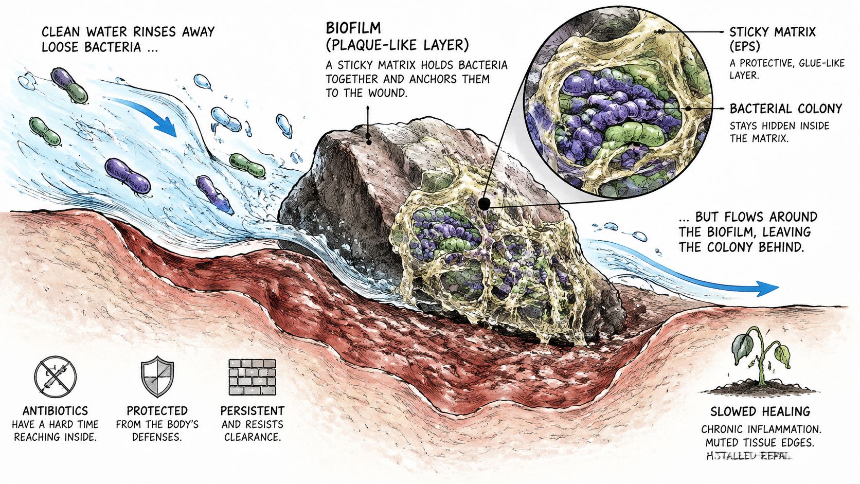
Think of the slippery coating attached to a wet rock. Rinsing may remove loose material while leaving the attached community behind. Biofilm in chronic wounds creates a similar practical problem, although its microscopic structure is more complex than the analogy suggests.
The illustration shows how microorganisms can remain embedded within a wound-associated matrix.
Protection changes the response to treatment
Biofilms are typically polymicrobial, meaning they contain different kinds of microorganisms rather than a single bacterial species. A review of wound biofilms and healing describes how their extracellular matrix and protected biological state increase tolerance to antimicrobial exposure compared with free-floating bacteria.
Tolerance isn't automatically the same as antibiotic resistance. Organisms living within biofilm may survive treatment because of their protected surroundings and altered activity, not necessarily because a laboratory test would classify them as genetically resistant.
This helps explain why prescribing another antibiotic isn't always the answer to a wound that remains open. Clinicians must identify what's preventing repair rather than assume persistent bacteria are the only problem.
Inflammation interferes with rebuilding tissue
A wound requires an early inflammatory response to control injury and contamination. When inflammation continues, however, increased protease activity and oxidative stress can interfere with tissue repair.
Proteases are enzymes that break down proteins. Excessive activity can disrupt the structural material the body needs to build new tissue, while oxidative stress can injure cells involved in healing.
Biofilm may also reform after disruption. A wound can therefore look better after treatment without sustaining measurable progress.
This accompanying video provides another way to visualize the topic.
Consider a hypothetical heel ulcer with a clean-looking base, a negative routine culture, and only a modest response to systemic antibiotics. If it barely contracts over successive visits, the team shouldn't treat the culture as proof that microbial involvement is absent.
Instead, clinicians should reassess tissue viability, recurring drainage, inflammation, pressure on the heel, and the response to previous treatment. They should also check whether the wound has the conditions it needs to rebuild tissue.
A stalled wound calls for reassessment of the whole wound environment. It doesn't identify a particular organism or prove that biofilm is the sole cause.
How to Spot Biofilm When a Wound Looks Clean
No single visible sign confirms biofilm. Slough, odor, and repeated breakdown can raise concern, but none is a dependable visual test.
A systematic review addressing biofilm-associated infection describes the diagnostic challenge: manifestations can be subtle, and laboratory confirmation remains limited. A wound that looks clean can still warrant investigation when its healing pattern is concerning.
Common wound findings and what they do and do not show
| Finding | Can suggest biofilm? | Why it is not conclusive |
|---|---|---|
| Slough or dead tissue | Possible | Nonspecific; also present in other stalled wounds |
| Odor | Possible | Can reflect other organisms or accumulated debris |
| Stalled healing or no size reduction | Possible | Poor perfusion, pressure, and offloading problems can also prevent progress |
| Recurrent breakdown | Possible | Shares causes with other wound complications |
| Negative routine swab | Biofilm isn't excluded | A surface swab may miss organisms embedded in tissue |
Slough should prompt assessment of tissue quality, not an automatic biofilm diagnosis. Odor needs interpretation alongside drainage, dressing condition, and other findings.
Failure to shrink and recurrent breakdown tell clinicians that the plan needs review. A negative swab provides information about the collected sample, not a complete map of the wound.
Separate microbial presence from infection
These terms describe different situations:
- Colonization means microorganisms are present without clinical evidence that they're causing infection.
- Local infection means microbial activity is causing harmful changes within the wound or its immediate surroundings.
- Spreading infection extends beyond the wound and needs prompt clinical attention.
- Suspected biofilm describes concern about an organized microbial community, not a separate visible stage of infection.
The distinction is particularly important for people with diabetes, pressure injuries, venous ulcers, or recurrent surgical wounds. Unnecessary antibiotics can cause harm, while missed infection can allow further tissue damage.
Compare progress, not just appearance
A clean-based wound and a wound containing slough may both fail to contract. Their appearance differs, but both need clinicians to investigate why healing has stopped.
Track trajectory, tissue quality, drainage, pain, odor, and undermining, which means tissue loss extending beneath the wound edge. Clinicians should interpret those findings alongside the patient's health risks and response to appropriate care, escalating when deterioration or repeated stalling persists.
How to Assess a Stalled Wound Step by Step
Assessment should produce a repeatable record, not just an impression that the wound looks better or worse. Consistent observations help clinicians distinguish temporary surface changes from meaningful healing.
The review of biofilm assessment and management describes how European guidance in 2014 favored biopsy or scraping after debridement for suspected bacterial biofilm, and how international consensus guidance in 2017 formalized biofilm-oriented care. The important shift was toward repeated assessment and targeted wound-bed management rather than a one-time prescription.
A repeatable clinical routine
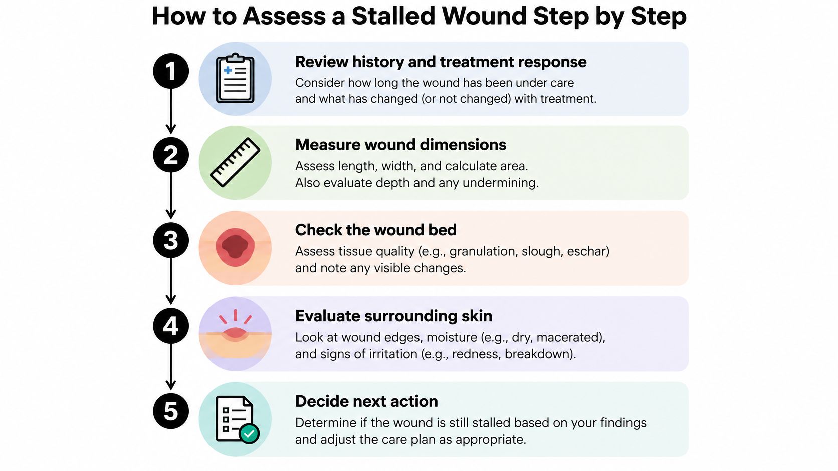
- Review the history and treatment response. Establish how long the wound has been under care, what treatments have been used, and what changed after each intervention. A common mistake is changing products without documenting why the previous approach didn't work.
- Measure consistently. Record wound area, depth, undermining, and drainage at planned intervals using the same method. Irregular measurements or different positioning can make an unchanged wound appear to improve.
- Examine the wound bed and surrounding skin. Check for devitalized tissue, edge changes, excess moisture, irritation, and tissue that may need removal. A clinician must determine whether debridement is safe, rather than assuming all dark or adherent tissue should be removed immediately.
- Choose sampling deliberately. When investigating suspected bacterial biofilm, biopsy or scraping after debridement may provide more representative material than a superficial swab. Swabbing accumulated surface debris can produce a result that doesn't adequately describe the tissue of concern.
- Interpret results within the clinical picture. Record laboratory findings beside wound measurements, symptoms, and treatment response. When considering molecular wound testing, distinguish identifying microorganisms from establishing their organization as biofilm or proving invasive infection.
- Compare the next assessment with the trend. Look for sustained contraction and improving tissue quality, not just a cleaner surface after a dressing change. Continuing an unchanged plan without checking its effect can delay a needed adjustment.
- Escalate when appropriate care produces limited improvement. Specialist review can reassess the diagnosis, wound environment, and suitability of further intervention. Referral shouldn't require caregivers to prove that biofilm is present first.
The infographic summarizes the main assessment checkpoints.
What caregivers can record between visits
Use photographs taken in similar lighting and from a consistent angle, with a measurement scale nearby if the care team recommends it. Keep a log of drainage, odor, discomfort, dressing saturation, and unexpected changes.
Don't probe the wound, scrape its surface, or attempt sharp debridement at home. Clinicians should measure depth and undermining and decide what tissue can safely be removed.
A useful wound log records what changed, when it changed, and what treatment was in place at the time.
Bring the actual product names and dressing-change routine to appointments. That detail is more useful than describing everything as an antibiotic dressing or wound cream.
How Biofilm Is Treated With Debridement and Topical Care
Repeated physical disruption combined with an appropriate topical strategy is the central management principle. Antimicrobial therapy alone doesn't address all the tissue conditions that allow an organized microbial community to persist.
A consensus review of biofilm management identifies effective debridement as a key intervention because it removes biofilm along with slough and devitalized tissue that provide attachment sites. The same guidance recognizes that residual biofilm may remain, which is why clinicians combine interventions rather than rely on removal alone.
What each treatment component adds
Biofilm management components and timing
| Component | Role | When it is used |
|---|---|---|
| Debridement, sharp or otherwise effective | Removes biofilm and supporting slough | Repeated at assessments where devitalized tissue is present and removal is safe |
| Antiseptic or antibiofilm dressing | Reduces residual organisms and limits reformation | Applied promptly after debridement when clinically appropriate |
| Cleansing or surfactant | Clears debris and helps loosen the matrix | At dressing changes according to the wound and clinical context |
| Systemic antibiotics | Treats invasive infection | For clinically diagnosed infection, not colonization alone |
Debridement isn't synonymous with an operation. Clinicians select a method according to tissue condition, circulation, wound location, discomfort, and what can safely be accomplished.
The topical component has a different job. After tissue removal, a selected antiseptic or antibiofilm-oriented dressing helps manage organisms left behind and supports control between assessments.
Cleansing removes loose material, while a surfactant can help loosen adherent debris and matrix. Patients shouldn't substitute household disinfectants or add products to a prescribed regimen without clinical guidance.
Systemic antibiotics belong in the plan when clinicians diagnose infection requiring them. Their role isn't to sterilize every colonized wound or compensate for unresolved pressure, dead tissue, or poor circulation.
Match treatment intensity to response
Consensus guidance describes an initial combination of maximally effective debridement and antibiofilm treatment, followed by stepped-down care as the wound improves. Stepping down means adjusting treatment to current need, not abandoning monitoring.
Regular debridement is associated with faster healing and wound-size reduction, but that association doesn't establish biofilm eradication as the independent reason a wound closes. Clinicians still need to judge whether the entire care plan is working.
For patients exploring advanced wound care options, the useful question is which unresolved barrier the proposed intervention addresses. A more advanced product isn't automatically a better fit for the wound.
Measure the wound's response
At planned reviews, clinicians should track:
- Area and depth, including whether tissue loss is decreasing.
- Undermining, including persistent spaces beneath wound edges.
- Exudate and pain, noting meaningful changes rather than isolated impressions.
- Granulation, the developing repair tissue within the wound bed.
Caregivers should report worsening between visits rather than wait for the next scheduled measurement. Limited response despite appropriate treatment is a reason to reconsider the diagnosis and care level, not continue the same dressing indefinitely.
What Biofilm Treatments Can and Cannot Promise
Will a silver dressing, antiseptic, or advanced product eradicate biofilm and close the wound? No product can responsibly promise both outcomes for every patient.
Conventional treatments can reduce surface biofilm burden, but complete eradication is uncommon. Successful management also requires attention to the conditions that prevented healing in the first place.
Microbial improvement isn't the same as closure
A 2026 systematic review and meta-analysis of randomized trials found no statistically significant difference in complete surgical-wound closure across two studies involving 284 participants. That finding applies to the studied surgical-wound comparisons, not every topical treatment or chronic wound.
It doesn't prove topical therapies are useless. It means a reduction measured in a laboratory or shortly after treatment shouldn't automatically become a claim of faster closure, fewer infections, or avoidance of procedures.
The illustration highlights the difference between reducing organisms at the surface and eliminating an established microbial community.
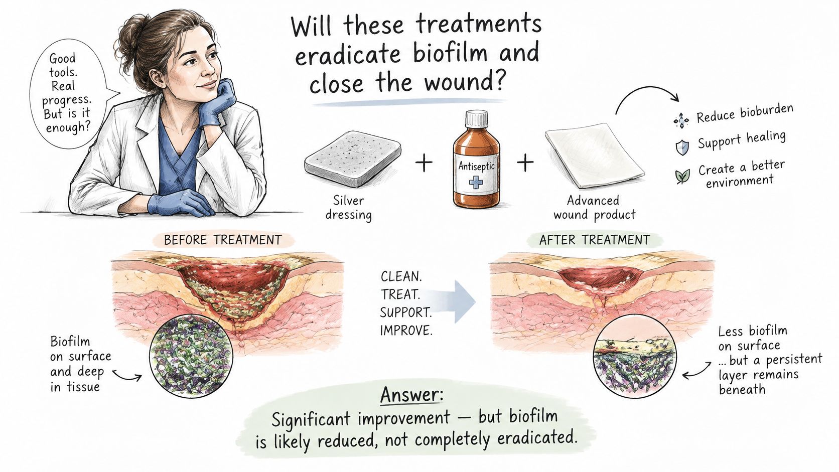
Treatment expectations: A cleaner-looking wound or lower microbial burden is an intermediate result. The patient still needs a plan for durable healing.
Promising options still need clinical proof
Developing approaches include bacteriophages, viruses that target bacteria, and quorum-sensing inhibitors, which interfere with microbial communication. Other approaches include matrix-degrading enzymes, electroceutical dressings, antifungal strategies, and nanotechnology.
These are developing options, not established replacements for wound-bed preparation and cause-directed care. A plausible mechanism doesn't establish safety, superiority, or a meaningful benefit for a particular patient.
Take these questions into the treatment discussion:
- Intended benefit. Is the treatment meant to control infection, improve the wound bed, manage drainage, or increase the likelihood of closure?
- Measurement. Which changes will demonstrate benefit over time?
- Change point. What finding would lead the team to stop, adjust, or escalate treatment?
Clear answers make uncertainty manageable. They also prevent repeated product changes from becoming a substitute for a coherent care plan.
Decide When a Stalled Wound Needs Specialist Care
A wound that stops shrinking, repeatedly reopens, or improves minimally despite appropriate evidence-based care deserves specialist review. Evaluation doesn't automatically mean surgery, and suspected biofilm alone isn't a reason to assume an operation is necessary.
The decision should rest on persistent tissue problems, healing response, and the patient's overall condition. Many stalled wounds can continue with conservative management once clinicians identify and address the missing barrier.
Findings that deserve a closer assessment
We list eight indicators that a wound may warrant surgical evaluation:
- No measurable size reduction despite ongoing care.
- Necrotic or dead tissue that requires assessment.
- Persistent undermining beneath the wound edge.
- Infected tissue or recurrent cellulitis.
- Exposed tendon or bone.
- Persistent slough tissue.
- Recurrent wound breakdown after apparent improvement.
- Minimal improvement despite appropriate evidence-based care.
An item on this checklist is a reason to request assessment, not a verdict that surgery is required. Our approach keeps conservative treatment as the foundation and coordinates recommendations with the patient's existing providers.
What specialty review involves
Through our wound escalation pathway, we review the history, wound, imaging, and treatment response virtually. A wound surgeon can examine the available images and the wound by video, with consultations usually offered within days.
A surgical wound-care nurse then validates the wound in person to confirm what was seen. This helps connect the initial review with the tissue findings needed for an appropriate treatment decision.
Before the visit, gather the wound log, dated photographs, measurements, previous culture results, and a list of treatments. Include information about pressure relief, offloading, compression when prescribed, and difficulties carrying out the plan at home.
Keep appropriate care going while arranging review
Continue the prescribed dressing and underlying-condition management unless the treating clinician changes the instructions. Don't stop effective conservative care because a referral has been requested.
Don't wait for a routine consultation if infection appears to be spreading or the patient becomes acutely unwell. Rapidly expanding redness, fever, severe worsening pain, or sudden deterioration needs prompt medical assessment.
When a checklist finding persists despite appropriate care, request specialist review and bring the treatment record. The purpose is to determine what the wound needs next, not to confirm a predetermined procedure.
---
The clinic offers virtual and in-person specialty evaluation for complex, stalled, and recurrent wounds, with surgical intervention considered only when clinically appropriate. If your wound isn't progressing despite appropriate care, visit Comprehensive Wound Services to request a review of its history, images, and treatment response alongside your existing care team.