Would you recognize a bone infection if it didn't hurt and didn't cause a fever? It is easy to assume you would. The mental image is vivid: a hot, swollen limb, shaking chills, pain bad enough to send someone to an emergency department that same night.
That picture is accurate for one form of the disease, and it's the form that dominates most lists of osteomyelitis signs and symptoms. It's also why so many quiet cases get missed. A bone infection can smolder for months with nothing more dramatic than a wound that won't close, and the person carrying it can feel entirely well.
What follows is a walking tour of how bone infections behave, why the same organism can produce a loud illness in one person and a whisper in another, and which patterns deserve a call to a clinician rather than another week of waiting.
## Table of Contents - Osteomyelitis Symptom Patterns at a Glance - Where the pattern settles - Why Fever and Pain Can Be Missing - Who gets the quiet version - Local Signs, Wound Appearance and Conditions That Mimic Bone Infection - What shifts concern toward bone - Two wounds that look nothing alike on the surface - What a clinician records at a wound check - How Doctors Confirm Bone Infection - The escalation that settles it - Red Flags That Need Prompt Medical Assessment - What to bring to the appointment - Book a Prompt Evaluation for Suspected Bone Infection
Osteomyelitis Symptom Patterns at a Glance
| Pattern | Typical Findings | Whole-Body Signs | Key Warning Sign |
|---|---|---|---|
| Acute osteomyelitis | Develops over days; localized bone pain that worsens quickly | Fever, malaise, raised white cell count | Pain escalating over days |
| Chronic osteomyelitis | Smolders for weeks or months; persistent or recurrent pain, swelling, warmth, tenderness | Few, and often none | Draining sinus or nonhealing wound over bone |
| Adult vertebral osteomyelitis | Persistent focal back or neck pain, local muscle tenderness | Fever in some cases only | Pain not improving with usual conservative care |
| Adult peripheral bone infection | Difficulty bearing weight, reduced limb movement, functional decline | Uncommon unless bacteria arrived through the bloodstream | Declining mobility in the affected limb |
| Diabetic foot with neuropathy | Pain may be mild or absent; wound progression, undermining, exposed bone | Minimal in most cases | Positive probe-to-bone test or exposed bone |
Presentation depends on two things: the type of infection and how long it has been running. Acute disease develops over days and is far more likely to produce whole-body inflammation. The American Academy of Family Physicians review of osteomyelitis lists fever, malaise, leukocytosis and rapidly worsening localized bone pain among its defining features. This is the version that gets diagnosed quickly, because it looks like infection everywhere else in the body.
Chronic osteomyelitis smolders for weeks or months with fewer systemic symptoms. The signs stay close to the bone: persistent or recurrent pain, swelling, redness, warmth, tenderness, a draining sinus, or a chronic nonhealing wound over the affected area. A fistulous tract connecting skin to bone is considered highly characteristic of chronic infection, and it's the finding that should end any debate about whether something deeper is going on.
Where the pattern settles
In adults, vertebral osteomyelitis usually shows up as persistent back or neck pain with local muscle tenderness, and fever appears only sometimes. Peripheral disease causes difficulty bearing weight, reduced limb movement, or a general loss of function in the limb. Pain can be mild or absent in people with peripheral neuropathy, including many with diabetes, so a comfortable limb proves nothing. Pediatric guidance draws a clean line at four weeks from the first symptom or sign, which separates acute osteomyelitis from the indolent form.
The reading rule: match the pattern and the duration first. Fever is a bonus clue, not the entry ticket.
Find the row that resembles what you're seeing. Escalating pain over a few days sits at one end. A wound that has drained intermittently for two months without a single fever sits at the other. They're the same disease, and they need the same conversation with a clinician, just on different timelines.
Why Fever and Pain Can Be Missing
Say a 62-year-old man with type 2 diabetes notices a blister on his heel. He offloads it, keeps it clean, and waits. Six weeks later the ulcer has stopped shrinking. He feels well. There's no fever, no chills, and the heel barely aches, because diabetic neuropathy has already dulled the nerves that would normally complain. His temperature is normal at every visit.
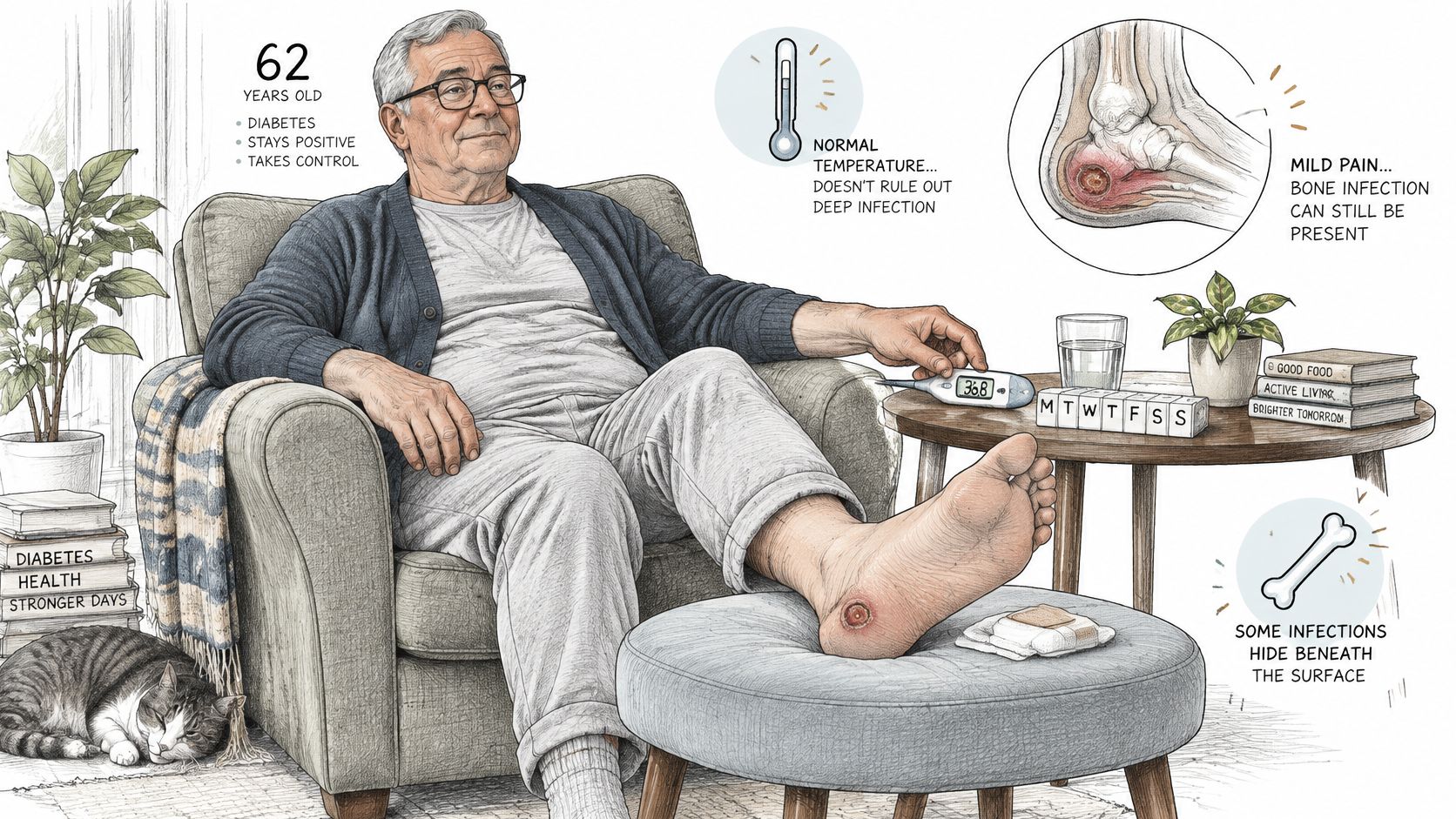
This is the case that gets missed, and it's why absence of fever does not rule out osteomyelitis, particularly in chronic, vertebral or wound-associated disease. The MSD Manual's guidance on osteomyelitis puts it plainly: chronic infection may present mainly with localized pain or tenderness and few constitutional symptoms.
Who gets the quiet version
Older adults, people with diabetes, anyone on immunosuppressive treatment and people living with neuropathy often mount a muted inflammatory response or struggle to describe pain that crept up slowly. Vertebral infection is the other quiet operator. Persistent, focal back pain that doesn't improve with the usual conservative care may be the only early sign; radiating pain, weakness, numbness or spinal-cord compression can follow later.
Symptoms alone can't confirm or exclude a bone infection. Imaging and microbiologic testing are what settle it.
The case series below show how wide that range runs. Fever was reported in most, but not all, of the children in each pediatric study, and the chronic jaw series was dominated by pain, swelling and visible bone rather than by systemic illness.
| Study and population | Reported frequencies |
|---|---|
| Pediatric osteomyelitis, 19 patients (BMC Pediatrics) | Pain 100.0%, tenderness 89.5%, limping 80.0%, fever 78.9%, decreased range of motion 73.7% |
| Pediatric fibular osteomyelitis, 13 patients (Frontiers in Surgery) | Fever 92.3%, bone pain 92.3%, local abscesses 76.9% |
| Chronic jaw osteomyelitis (Journal of Clinical Medicine) | Pain 92.5%, swelling 81.1%, bone exposure 66.0%, fistula formation 43.4%, limited mouth opening 34.9% |
All three are small series, so read the percentages as a picture of variation, not as a risk estimate for any individual person.
Local Signs, Wound Appearance and Conditions That Mimic Bone Infection
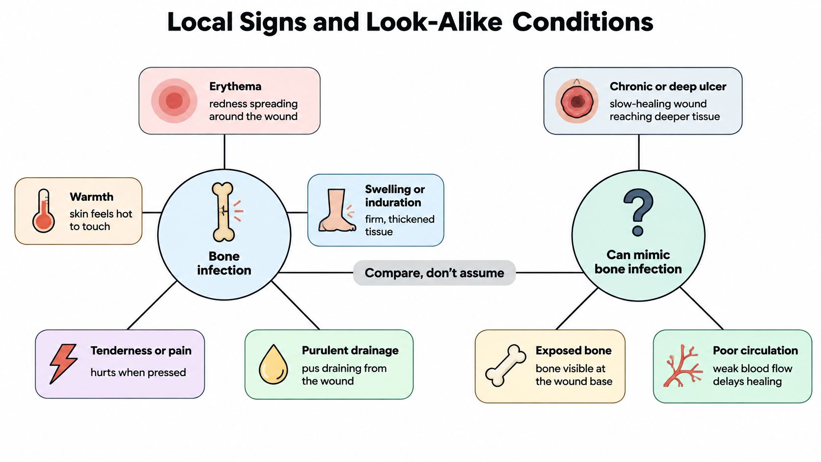
Five local features come first at any wound check: erythema (redness), warmth, swelling or induration, tenderness or pain, and purulent drainage. Together they point to infection in the tissue around the wound. Whether that infection has reached bone is a separate question with separate evidence. The most useful way to read osteomyelitis signs and symptoms at the wound bed is as a pattern rather than a checklist, and the IWGDF/IDSA practical guidelines on diabetic foot infection frame it exactly that way.
What shifts concern toward bone
A chronic or deep ulcer, exposed bone, or a positive sterile probe-to-bone test all raise the possibility that infection has crossed into bone. The probe result is weaker evidence than it looks. Its meaning depends on how deep the ulcer is, where it sits, how common osteomyelitis is in that patient group, and whether anything else supports the finding. Current guidance recommends pairing probe-to-bone testing with plain radiographs and an inflammatory marker such as ESR, CRP or procalcitonin before drawing conclusions.
Raised markers strengthen suspicion without confirming anything. Cellulitis, trauma, gout, Charcot neuroarthropathy and a recent operation can all push inflammatory markers upward.
Two wounds that look nothing alike on the surface
A surgical wound with bright, spreading redness that settles once the soft-tissue infection is treated is a common story, and a reassuring one. A deep puncture wound from a thorn or a pet bite can look almost calm on the surface while the person describes a fixed, boring pain in the bone underneath that hasn't changed in three weeks. The first behaves like soft tissue. The second behaves like bone, and appearance alone won't separate them.
That's the trap. Redness, drainage, odor or visible exposed tissue may reflect soft-tissue infection, inflammation, poor circulation or simple colonization rather than osteomyelitis, as Mayo Clinic's guidance on diagnosing osteomyelitis notes. The reverse holds too: bone can be infected with very little surface inflammation. An early X-ray won't rescue the assessment either, because visible bone damage may not appear until the infection has been present for weeks.
Progression is the signal that matters most. A wound that deepens, undermines further, or drains again after drying up is telling you something a single red photograph cannot. Complex or repeatedly reopening wounds deserve advanced wound care review rather than another dressing change.
What a clinician records at a wound check
- Measurements: length, width and depth in centimeters at every visit, so the trend exists in numbers rather than in memory.
- Depth and undermining: whether a probe disappears under the wound edge, and in which direction.
- Probe-to-bone: recorded with its result and whatever supported it, never as a standalone yes or no.
- Circulation and sensation: pulses, capillary refill and a monofilament check, because poor blood flow changes healing and neuropathy changes reporting.
- Dated photographs: taken the same way each time, with a ruler in frame.
- Timeline: when the wound opened, what has been tried, and whether it has ever closed and then reopened.
Ask which of these were recorded at your last visit. If the answer is none of them, the trend you need doesn't exist yet.
How Doctors Confirm Bone Infection
Diagnosis runs as a sequence of narrowing questions, and each test answers a different one.
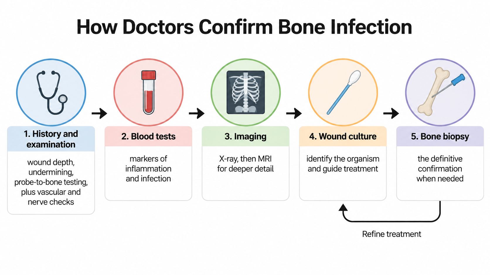
Examination comes first, and it's more structured than it sounds. The clinician measures the wound, checks for undermining, probes for bone in a deep ulcer, and assesses pulses and sensation in the limb. A history of previous surgery, an open fracture, a recent joint replacement or a distant infection all shift the pretest probability before any image is taken.
Blood work follows. Inflammatory markers such as ESR, CRP or procalcitonin rise during infection and are useful for tracking a response, but they can't distinguish bone from soft tissue, and plenty of other conditions raise them. Plain radiographs come next as a baseline. Cortical destruction, periosteal reaction, sclerosis, sequestrum and soft-tissue gas are all visible findings, though early disease often isn't.
The escalation that settles it
MRI is the escalation test when examination, X-rays and labs leave the question open. Osteomyelitis typically produces confluent low T1 marrow signal with high fluid-sensitive signal and enhancement, and sinus tracts, intraosseous abscesses or sequestra add anatomical evidence. The IWGDF guideline on diagnosing diabetic foot infection describes MRI sensitivity around 90% and specificity near 80%. A normal scan makes infection unlikely in many settings, though postoperative change, fracture and Charcot arthropathy can pull specificity down. CT steps in when MRI is unavailable or when surgical planning needs finer bone anatomy.
When imaging and clinical findings still disagree, bone biopsy with histology and culture is the most definitive method for identifying infection and guiding antibiotic choice. Molecular techniques applied to molecular testing of a properly collected sample can also name an organism when routine cultures come back negative.
This short walkthrough covers the same diagnostic logic in visual form.
| Test | What It Shows | Main Limitation |
|---|---|---|
| Inflammatory markers (ESR, CRP, procalcitonin) | Raised levels strengthen suspicion | Nonspecific; cellulitis, trauma, gout or Charcot can raise them |
| Probe-to-bone test | Sterile probe contacts bone in a deep ulcer | Not diagnostic alone; depends on ulcer depth and location |
| Plain X-ray | Baseline view of cortical destruction, periosteal reaction, sequestrum, soft-tissue gas | Early disease can be radiographically occult |
| MRI | Marrow involvement, sinus tracts, intraosseous abscesses; sensitivity around 90%, specificity near 80% | Postoperative change, fracture or Charcot can reduce specificity |
| CT | Detailed bone anatomy when MRI is unavailable | Less sensitive for early marrow change |
| Bone biopsy with culture | Histology and the causative organism to guide antibiotics | Invasive; used when findings stay discordant |
Red Flags That Need Prompt Medical Assessment
The useful question isn't whether a symptom is an emergency. It's how quickly it needs a clinician's eyes on it.
| Finding | Timing | Action |
|---|---|---|
| Progressive weakness or numbness with spinal pain | Immediately | Emergency care |
| Fever with bone pain, swelling or redness over bone | Same day | Urgent medical evaluation |
| Rapidly worsening localized bone pain | Same day | Urgent medical evaluation |
| Persistent focal bone or back pain without fever | Within days | Medical assessment |
| Draining sinus, recurrent wound breakdown or exposed bone | Within days | Prompt specialist review |
| Diabetic foot wound with no improvement after appropriate care | Within days | Prompt specialist review |
| Difficulty bearing weight or reduced limb movement | Within days | Medical assessment |
Two rows above carry most of the urgency. Progressive weakness or numbness alongside spinal pain can signal cord compression, which is a same-hour problem, not a same-week one. Fever with pain, swelling or redness over a bone points toward acute infection and needs evaluation the same day.
What to bring to the appointment
- A symptom timeline: when the pain or wound started, whether it has improved, and what makes it worse.
- Wound records: measurements or dated photos, including any stretch when it closed and then broke down again.
- Prior imaging and reports: the actual discs or files where possible, not just a one-line summary.
- Medication history: antibiotics already taken, steroids, immunosuppressants and diabetes medication.
- Recent glucose readings: for anyone with diabetes, the trend across the past few weeks.
Pain or a wound that outlasts the expected recovery window is a reason to be seen, not a reason to keep watching.
Function that doesn't come back counts as a finding too. A limb you can't bear weight on after an injury or an infection belongs on the same list. A structured escalation pathway moves a stalled wound to the right level of review without waiting for a crisis to force it.
Book a Prompt Evaluation for Suspected Bone Infection
A wound over bone that has stalled, drained for weeks, or reopened after closing deserves medical evaluation. The pattern and duration of symptoms matter more than whether fever is present.
When arranging an appointment, ask what each test is meant to clarify: whether inflammatory markers, probe-to-bone testing, imaging, or a bone biopsy may be appropriate. Ask how healing will be tracked, such as with measurements or dated photographs, and what changes should prompt reassessment.
For a stalled or recurrent wound, Wound Services offers a surgical wound evaluation pathway. Conservative care remains the foundation. If progress stops, a wound surgeon can review the history, images, imaging, treatment response, and wound by video, usually within days. An evaluation does not automatically lead to surgery. Primary care and hospital-based wound services are also options.
Continue appropriate wound care, such as dressings, offloading, compression, nutrition, and treatment of underlying conditions. Surgery may be considered when healing stalls or bone is involved, if clinically appropriate.
The wound team evaluates complex, stalled, and surgically appropriate wounds, including wounds with suspected deep infection or bone involvement. If a wound has stopped shrinking, repeatedly reopens, or exposes bone, request a virtual consultation so a wound surgeon can review the available information and discuss next steps. Visit Comprehensive Wound Services to start the consultation process.