
Why does a wound smaller than a coin sometimes outlast one twice its width?
The opening is the doorway. Wound tunneling is the narrow channel that runs from that visible bed into surrounding or deeper tissue, and the doorway says little about how far the corridor goes. Measuring only the skin opening underestimates the damage, because the actual size sits out of sight.
In full-thickness pressure ulcers, tunneling is less common than undermining. A quieter rate does not shrink the problem inside one wound. Fluid can sit in the passage. The packed dressing has to follow the channel, not stop at the entrance. Closure counts when that passage shortens, not when the surface looks calmer.
A modest opening can still be the wrong place to stop looking.
## Table of Contents - What Wound Tunneling Is and How It Differs from Undermining - Why a Small Opening Can Hide a Large Problem - How Clinicians Measure and Probe a Tunnel - The probing sequence, step by step - What Causes Tunneling and Where Treatment Starts - Start with the cause, not the dressing - Signs that change the plan - Tunneling in Real Wounds Across Three Common Cases - Three scenarios and what each one hid - What a tract tends to be holding - When to Escalate, Follow Up, and Who to Involve - Who to involve, and what follow-up looks like - Signs to report promptly
What Wound Tunneling Is and How It Differs from Undermining
Think of a burrow dug under a lawn. One small hole at the grass line, and a passage running off in a direction nothing above the soil hints at. A wound tunnel behaves the same way, which is why looking at the entrance tells you so little.
If you've looked up what wound tunneling is and come away with a picture of a narrow passage, you have the shape right. The word clinicians add is directional. A tunnel goes somewhere. Undermining is tissue separation beneath the wound edge, and it usually spreads around that edge instead of running off on one bearing.
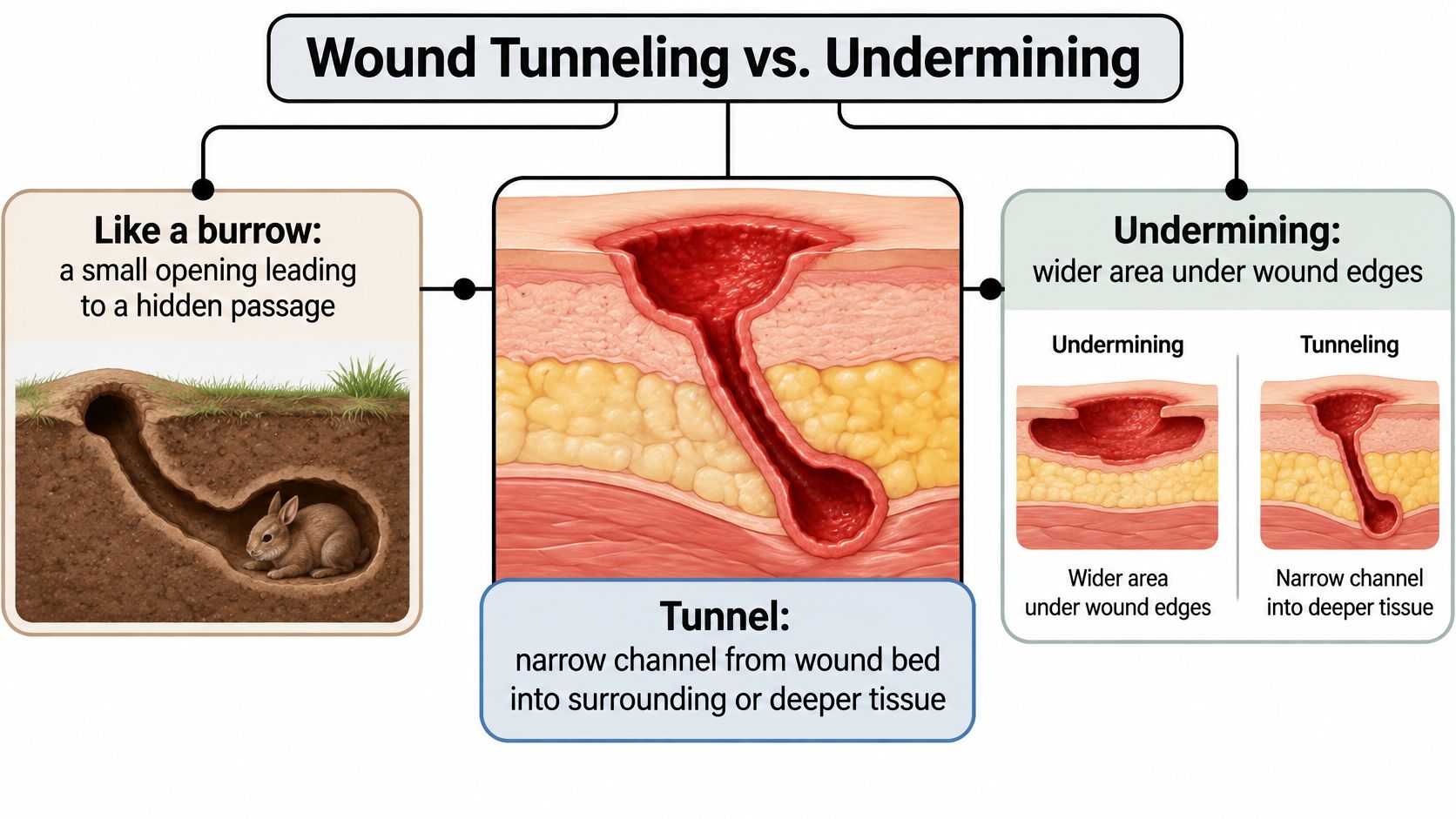
The three terms get used interchangeably in conversation, and they shouldn't be.
| Feature | Wound tunneling | Undermining | Sinus tract |
|---|---|---|---|
| Shape | Narrow channel from the wound bed | Broad separation beneath the wound edge | Narrow tract that may end in a drainage opening |
| Direction | Directional, tract-like | Often spreads around the edge | Usually runs toward the surface |
| Documented as | Depth, length and clock-face direction | Depth and clock-face position of the separated tissue | Tract depth and number of openings |
| Share in pressure ulcers | 5.2% of full-thickness wounds (2026 registry analysis) | 15.6% of wounds in the same analysis | Not reported separately in that analysis |
A 2026 registry-based analysis of about 32,000 documented pressure ulcers put numbers on the difference: tunneling in 764 of 14,700 full-thickness wounds (5.2%) and undermining in 2,293 (15.6%). The same analysis described tunneling as typically short and directionally clustered relative to the wound bed, and it matches assessment guidance that asks for location, stage, surface dimensions, depth, sinus tracts, undermining, tunneling, exudate, necrotic tissue, granulation tissue and epithelialization at the first assessment.
Tunneling isn't confined to one wound type. It appears in pressure injuries, in surgical wounds that have opened, in diabetic wounds, and in other deep or infected wounds, which is why the same questions get asked whether the patient is at home, in a nursing facility, or in a hospital bed.
A small surface opening does not mean a small wound. It means the part you can see is the smallest part of it.
That's the practical lesson. A wound with a modest opening that has a tract behind it deserves careful probing, clear documentation, and repeat assessment, because the surface measurement alone will keep telling you everything is fine.
Why a Small Opening Can Hide a Large Problem
A tunnel isn't a surface feature. Its presence usually means the injury has reached full thickness, and full-thickness damage is what lets a tract conceal devitalized tissue, infection, or a route to structures far below the skin.
Staging makes that concrete. In a stage 4 pressure injury, full-thickness skin and tissue loss can expose or allow direct palpation of fascia, muscle, tendon, ligament, cartilage or bone, and the 2022 spinal cord injury pressure ulcer guidelines list tunneling and undermining as features that commonly occur in those advanced wounds. A stage 3 injury can tunnel too, though exposed fascia, muscle, tendon, cartilage and bone aren't part of that picture unless the wound turns out to be more advanced or unstageable.
This is a relatively recent way of thinking. Pressure-injury classification used to describe surface sores. It now asks for depth and for subsurface characteristics, sinus tracts, undermining and tunneling included, alongside the visible dimensions.
Why the surface can look better while the wound isn't: a tract holds drainage and dead tissue at the far end of the passage, so the edges can close over a space that never actually filled in.
Retention is the mechanical reason this matters. A tunnel can trap exudate or necrotic material and keep the wound from closing even when the top looks like it's turning a corner. Watch for a wound that stops shrinking, reopens repeatedly, drains persistently, smells, hurts more, or shows spreading inflammation around the rim. Each of those signals a reason to reassess rather than reassure.
Deep stage 3 wounds with undermining tracks, and stage 4 wounds that fail standard care, may need surgical referral under the same guidance. Tunneling is a marker of wound complexity, not a footnote in a measurement chart, and the last section covers who to involve and when.
How Clinicians Measure and Probe a Tunnel
Length and width are the first two numbers anyone writes down, and they're the two that mislead most once a tract is present. The external opening can be far smaller than the passage behind it, so a real assessment adds depth along the tract and the direction it runs.
| Element | What to record | Why it matters |
|---|---|---|
| Tunnel depth | Length from the wound base to first resistance, in centimetres | Shows the hidden extent of the wound |
| Direction | Clock-face position, with 12 o'clock toward the head | Tells the next clinician where the passage leads |
| Exudate and wound bed | Drainage, slough, necrosis and granulation | Flags infection and devitalized tissue |
| Change over time | Repeat measurements at each review | Separates healing from progression |
A nursing wound-assessment chapter in the LOUIS Pressbooks open textbook describes the technique plainly: a sterile, blunt probe or cotton-tipped applicator advanced gently from the wound base until resistance is met. Force is never used, because pushing can injure viable tissue or create a false passage that later gets measured as if it were real. Direction is charted on a clock face with 12 o'clock pointing toward the patient's head, and the reading is repeated at each review so contraction can be told apart from progression.
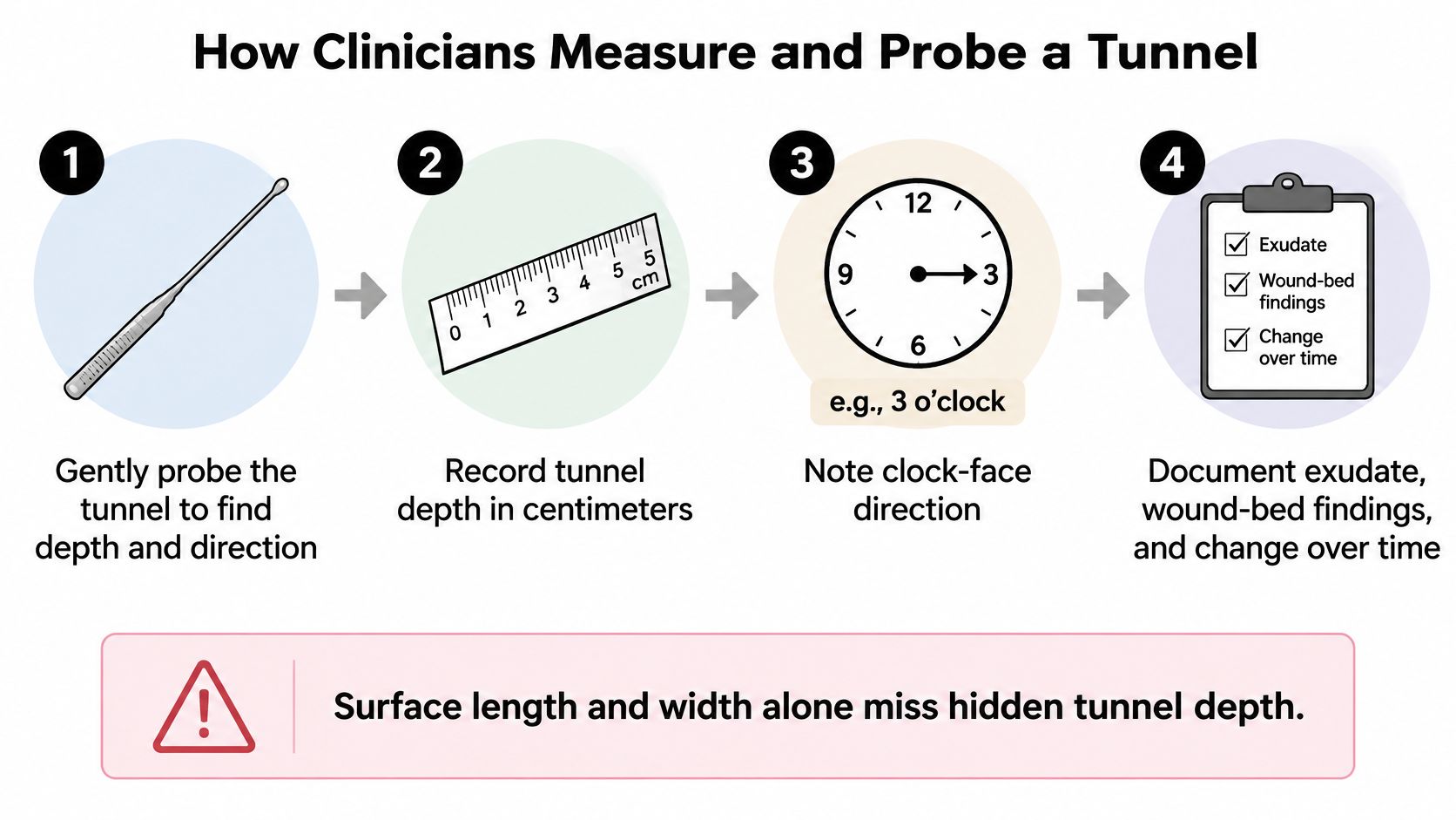
Get those four elements recorded and the tract stops being a mystery and starts being a trend line.
The probing sequence, step by step
- Expose the whole wound and a margin of skin around it. Position the patient so nothing folds over the opening. A tract that runs toward the hip or the natal cleft is invisible if the patient is lying in a way that compresses it.
- Choose a blunt instrument, not a sharp one. A sterile, blunt probe or a cotton-tipped applicator is the tool. Sharp instruments belong in a procedure, not a bedside assessment.
- Advance from the base until you meet the first real stop. Stop there. The resistance you feel is the end of the tract or the wall of it, and pushing past either one gives you a false number and a tissue injury.
- Record length and clock position in the same note. "3 cm at 3 o'clock" is useful. "Deep" is not, and neither is a number with no direction attached.
- Repeat on a schedule, weekly or sooner if something changed. Consistent serial measurements are how a care team tells a wound that's filling in from one that's extending.
Photographs help families and clinicians see the same thing, and a walkthrough of photographing and measuring wounds over time covers that side of the record. In our own workflow we review the history, wound, imaging and treatment response virtually first, then a surgical wound care nurse validates the wound in person to confirm what was seen. The bedside measurement and the specialist read should agree.
What Causes Tunneling and Where Treatment Starts
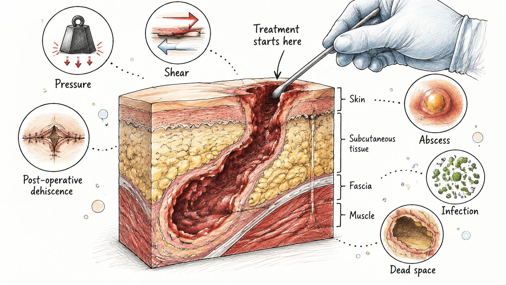
A tunnel is a cavity problem, not a dressing problem, and superficial dressing changes alone don't manage an irregular cavity. That's the reasoning behind the 2023 Wound Healing Society pressure ulcer guideline, which recommends that irregular wound extensions forming sinuses or cavities be explored, unroofed and treated, because a closed or narrow tract can hide nonviable tissue and infection.
Start with the cause, not the dressing
The usual drivers are pressure, shear, a surgical wound that has opened, an abscess, infection, or dead space that persists because nothing fills it. Treatment begins by naming which of those is at work, then removing the ongoing barriers: pressure the tissue can't tolerate, moisture that macerates the edges, ischemia, malnutrition, and uncontrolled diabetes.
Fix the barrier and the tract has a chance to fill. Skip it and even a well-packed tunnel keeps reforming in the same place.
Signs that change the plan
Infection is the complication that most often forces escalation. Worsening pain, purulent drainage, spreading redness, warmth, induration, new necrosis, foul odour, or a wound that stops improving all point the same way, and each warrants prompt clinical review rather than a dressing change.
A superficial swab is a weak guide to a deep infection. Tissue or bone sampling is preferred when the diagnosis is unclear. When a tract runs toward bone, or osteomyelitis is suspected, imaging becomes part of the workup, and established cavity-wound guidance names CT or MRI among the studies used to see what probing cannot reach.
The short video below gives a visual sense of how deep these wounds run.
Negative-pressure wound therapy has a real place here for selected chronic stage III or IV pressure ulcers. What it doesn't do is replace debridement, source control, pressure redistribution, or a specialist review when the tract keeps reforming. Think of it as one tool in an advanced wound care plan, not the plan itself.
Tunneling in Real Wounds Across Three Common Cases
Three scenarios show how differently a tract can present and why the same finding leads to different plans. These are illustrative examples, not accounts of individual patients.
Three scenarios and what each one hid
A sacral pressure injury with a small opening. A caregiver reports a shallow-looking sore over the tailbone, smaller than a coin, that hasn't changed in six weeks. Probing finds a tract running several centimetres toward the hip at 4 o'clock. That single measurement changes the stage, the support surface, the turning schedule, and the referral.
A surgical wound that keeps reopening. An abdominal incision separates, gets packed, closes, then breaks down again two months later. Dressings are appropriate and the home nurse is diligent. What's missing is the pocket at the base of the tract holding drainage, which keeps the surface from ever sealing for good.
A diabetic foot wound with a deep tract. The visible ulcer is unremarkable, but the foot is swollen, the pain is worse, and there's a new smell. Those clinical signs, not the surface size, drive the decision to escalate the same day.
What a tract tends to be holding
A 2025 case series of 89 chronic wounds with sinus tracts followed over four years, published in Wounds, found foreign bodies inside the tunnel in 8 cases (8.99%) and necrotic tissue in 32 (35.95%).
That second figure is the one to sit with. Roughly a third of those tracts contained dead tissue that no surface inspection would have found, and material of any kind sitting at the end of a blind passage keeps a wound open. When a wound plateaus, the tract is one of the first places to look and one of the easiest to miss.
When to Escalate, Follow Up, and Who to Involve
A tunnel can travel well past the small opening you can see. Judging the wound by that opening is like judging a house by the front door: the rooms behind it determine its size. Escalation is how a care team responds when that hidden length has a structural problem dressings cannot reach.
The people already around the patient do most of the work. That is the primary provider, home health staff, or skilled nursing staff who see the wound every week. A specialist joins that group rather than replacing it. Recommendations go back to the original team so care continues where the patient lives.
Wound Services uses eight indicators to flag a wound that may deserve surgical evaluation. They are concrete things to bring to any care team:
- No measurable size reduction after a reasonable course of appropriate care.
- Necrotic or dead tissue that persists despite debridement.
- Persistent undermining.
- Infected tissue or recurrent cellulitis in the same area.
- Exposed tendon or bone, which changes the wound from complex to urgent.
- Persistent slough tissue.
- Recurrent wound breakdown.
- Minimal improvement despite appropriate evidence-based care.
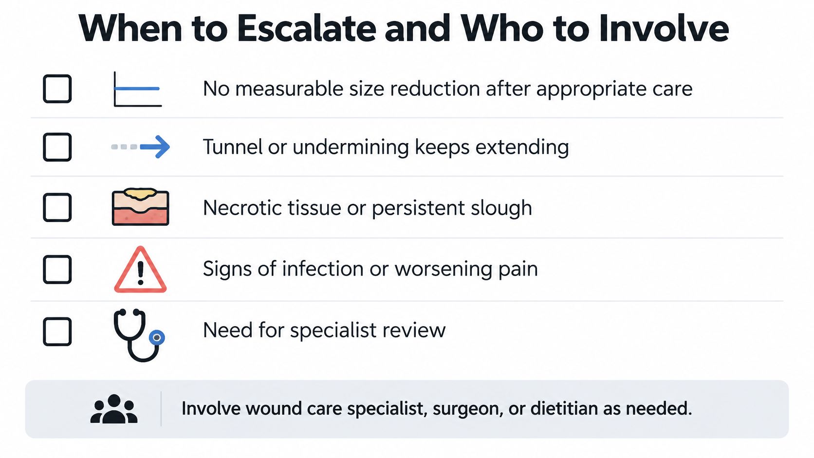
Who to involve, and what follow-up looks like
Follow-up after any specialist review uses the same measurement habits, repeated. Depth, direction, and the number of tracts get tracked from visit to visit until the wound closes. A tract that keeps extending on those repeat measurements belongs on the list above, because the surface can stay small while the passage grows. Virtual follow-up keeps that tracking going between appointments so a plateau gets caught early.
Signs to report promptly
Beyond the infection picture, three changes deserve a same-day call: bleeding from the wound or the tract, a tunnel that measures deeper than it did last time, and no measurable change after a stretch of appropriate care. Each one is a reason for a prompt conversation with the care team.
Three questions worth writing down before the next visit: Can you document the depth, direction and number of tracts at each assessment? Would imaging or a tissue sample answer something probing can't? And given how long this has been open, is a specialist evaluation appropriate?
Wound Services evaluates complex and stalled wounds virtually or in person and works alongside the team already in place. The escalation pathway for surgical wound evaluation explains how that handoff works, and an evaluation does not commit anyone to surgery. Before the first appointment, gather the wound history, recent photographs with dates, imaging reports, and treatment notes. The timeline of what has been tried is often the most useful thing in the room.
The Wound Services team reviews complex and stalled wounds, including wounds with tunneling or persistent undermining, through virtual or in-person specialty evaluation and, when clinically appropriate, surgery at its own ambulatory surgical center. If a wound has stopped shrinking, keeps reopening, or measures deeper than it did a month ago, that is the moment to get a specialist read on it. Visit Comprehensive Wound Services to start an evaluation or refer a patient.