Your parent came home from the hospital last week with a wound on the lower back. The home-health nurse lifts the dressing, and the opening looks smaller than you expected, maybe the width of a thumbnail. At the base, though, there's a pale, firm surface that doesn't look like skin, and the nurse says the wound is stage 4.
That moment is where many families get stuck. A stage 4 pressure injury is not a comment on how dramatic the wound looks from across the room. It's a description of how deep the damage goes and which structures the wound has reached. Once you understand that distinction, the diagnosis stops being a label and becomes a set of decisions.
## Table of Contents - How to Recognize a Stage 4 Pressure Injury - Why depth matters more than size - What to Measure When Assessing Depth and Extent - A checklist you can copy into a log - Why a baseline map matters - When Bone Exposure Signals Infection - What source control means - How Stage 4 Care Is Planned Step by Step - What progress looks like - When Surgery Is Considered and When It Is Not - Who reconstruction is not for - The four-step pathway - Complications and Long-Term Outlook for Stage 4 - What the numbers do and do not prove - Next Steps for a Stage 4 Wound
How to Recognize a Stage 4 Pressure Injury
A stage 4 pressure injury is full-thickness loss of skin and tissue with exposed or directly palpable fascia, muscle, tendon, ligament, cartilage, or bone. The NPIAP staging poster places it as the most extensive category in the staging system.
| Stage | Tissue depth | Visible signs |
|---|---|---|
| Stage 1 | Intact skin with non-blanchable redness | Skin is unbroken; redness does not fade when pressed |
| Stage 2 | Partial-thickness loss into the dermis | Shallow open area or blister |
| Stage 3 | Full-thickness loss through the subcutaneous tissue | Fat may be visible; fascia, muscle, tendon and bone are not exposed |
| Stage 4 | Full-thickness loss with exposed or palpable fascia, muscle, tendon, ligament, cartilage or bone | Slough or eschar may be present; undermining and tunneling are common |
| Unstageable | Depth cannot be confirmed because slough or eschar covers the base | Wound base hidden until cleaned or assessed |
Read the rows from top to bottom and you're following the damage through the layers of the body. Stage 1 stays in the skin. Stage 2 reaches the dermis. Stage 3 passes through the subcutaneous fat. Stage 4 goes beyond fat and reaches structures that normally sit under muscle or bone.
Why depth matters more than size
Depth varies by body site. Over the sacrum or heel, where there's little subcutaneous tissue, bone can be exposed quickly. Over the buttock or thigh, a wound can destroy a large amount of soft tissue before bone becomes visible. That's why a small opening over a bony prominence can be stage 4 while a wider, shallow scrape is not.
Slough is yellow, tan or grey dead tissue that clings to the wound bed. Eschar is dark, leathery dead tissue. Rolled edges, undermining and tunneling are common in stage 4. Undermining means a lip of tissue loss extends under skin that looks intact. Tunneling means a narrow channel runs away from the main opening.
In April 2016, NPIAP replaced pressure ulcer with pressure injury. The change recognizes that pressure-related damage can occur even when skin remains intact. You'll still see pressure ulcer in charts, billing records and older discharge summaries, so both terms describe the same condition. The term pressure ulcer stage 4 remains common in clinical and administrative use, but the staging definition is the same.
Stage 4 describes anatomical severity, not wound size, duration or infection status. That distinction matters for documentation, escalation, outcome measurement and communication between hospitals, nursing facilities, home-health clinicians, patients and caregivers.
The rule that prevents a common error: if slough or eschar completely hides the wound base, the injury is unstageable until the base can be assessed. It is not stage 4 yet.
What to Measure When Assessing Depth and Extent
A measuring tape tells you how wide the opening is. It cannot tell you how far the damage travels under skin that still looks whole. Two wounds with the same surface opening can hold very different amounts of lost tissue, so a surface measurement alone can substantially underestimate a stage 4 wound's true extent.
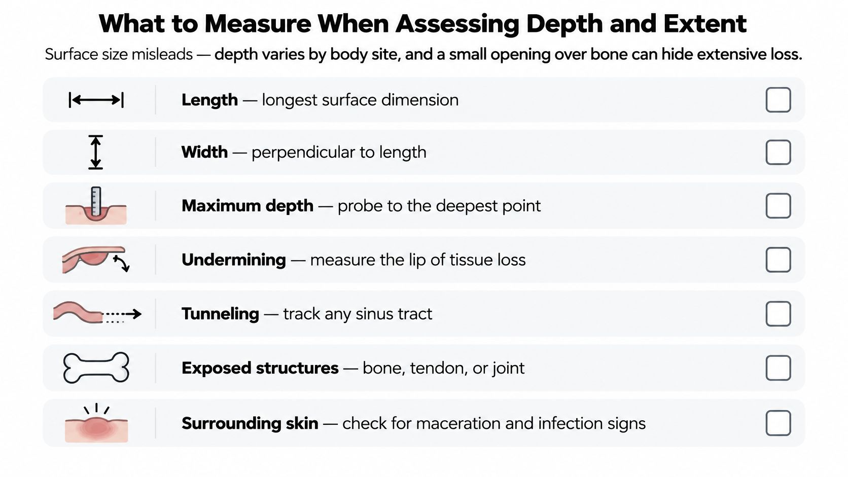
This visual shows the measurements that belong in a wound assessment.
A checklist you can copy into a log
- Length and width: record the longest surface dimension and the line perpendicular to it.
- Maximum depth: a soft probe is placed gently at the deepest reachable point, and that distance is recorded.
- Undermining by clock position: measure how far the lip of tissue loss extends and name where it sits.
- Tunneling: track any sinus tract, including its direction and depth.
- Exposed structures: note bone, tendon, joint or other deep tissue you can see or feel.
- Tissue type: describe how much of the bed is slough, eschar, granulation or new epithelium.
- Drainage: record amount, color and consistency after cleansing.
- Odor: check after cleansing, not before, so the dressing's smell does not confuse the finding.
- Periwound skin: look for maceration, redness, warmth or firmness around the edge.
- Pain: ask about pain before, during and after care, because a change in pain can signal infection or a deepening injury.
- Serial change: compare each entry with the last assessment, not with a general impression.
The clock face is a shared language. By convention, 12 o'clock points toward the patient's head and 6 o'clock toward the feet. If undermining runs from 2 to 6 o'clock, write that down. A note that says only that undermining is present doesn't tell the next clinician where the dead space sits or whether it grew.
Why a baseline map matters
Record a baseline wound map at the first full assessment: measurements, clock positions, tissue types, drainage, odor, periwound skin and pain. Take the photograph from the same angle, with the same lighting and the same measurement reference in frame each time. Every later note should compare with that map, not with memory.
The payoff is trend detection. A wound can hold the same surface opening for weeks while undermining widens underneath, and only a series of clock-position readings will catch that. The reverse is also true: shrinking depth at a steady opening is real progress even when the wound still looks dramatic.
If bone or tendon is visible or can be felt with a probe, record it in the map and escalate it. That finding moves the wound out of routine measurement and into a structured evaluation rather than a routine dressing change.
This short video shows how a clinician moves through the assessment sequence.
Three actions for this week:
- Start the log today. Write the date, measurements, clock positions and a photograph note on one page.
- Use the same fields every time. Consistency is what makes a trend visible.
- Bring the trend to the next visit. A series of readings tells the team more than a single dramatic photo.
When Bone Exposure Signals Infection
A stage 4 injury carries a high risk of deep infection because tissue loss can create a direct route from contaminated soft tissue to muscle and bone. The Wounds UK best-practice summary draws a useful distinction between superficial cortical involvement and medullary osteomyelitis, and describes approximately 2 weeks of antibiotic therapy for more superficial disease and 4 to 6 weeks when medullary bone is affected. Treatment is individualized, and extending antibiotics beyond 6 weeks is not evidence-based.
Cortical bone is the hard outer shell. Medullary bone is the inner marrow space. Infection that stays in the cortex is a different problem from infection that reaches the marrow, which is why the duration of treatment changes.
What source control means
Antibiotics without source control are like watering a plant whose root is still rotting. The medicine may reach the edge of the problem, but dead and infected tissue keeps feeding it. Source control means drainage of an abscess, removal of necrotic tissue, and soft-tissue or selected bone debridement when a specialist judges it necessary.
Exposed bone alone does not prove infection. Clinicians integrate the clinical picture, inflammatory markers, imaging and, when appropriate, bone sampling before committing to a long antibiotic course. A clean-looking bone surface in a stable patient is not the same finding as a draining wound with new pain and fever.
Report these signs promptly:
- Fever or chills that don't have another explanation.
- Spreading redness or warmth beyond the wound edge.
- New or worsening pain at the site.
- Foul drainage or a change in drainage color.
- Confusion or a change in alertness, especially in an older adult.
Lack of measurable improvement despite adequate off-loading and local care should prompt reassessment. The team should look for ongoing pressure, poor perfusion, malnutrition, hidden infection, inadequate debridement or a need for procedural management. Adding a stronger topical product before checking those causes usually delays the right treatment.
A practical trigger to escalate: a wound that is clean but not contracting after good pressure relief deserves a fresh look at the whole plan, not another dressing.
How Stage 4 Care Is Planned Step by Step
Treatment for a stage 4 wound is a bundle, not a product. Our advanced wound care service is built around that foundation: dressings, offloading, nutrition, pressure relief and treatment of underlying conditions. The plan works only when the basics are running every day, not just on visit days.
This visual shows the core sequence.
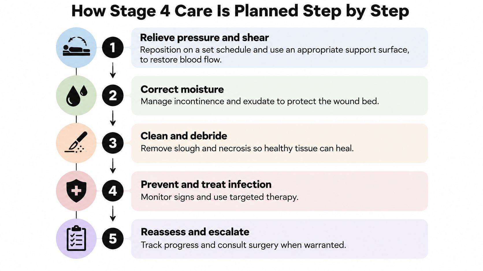
- Remove pressure and shear. Reposition on a set schedule and use an appropriate support surface. Pressure closes small blood vessels, so relief is what allows tissue to recover. The common slip is turning a patient onto the sore hip because it's the easier side.
- Correct moisture and friction. Manage incontinence, sweat and sliding in bed. Wet skin breaks down faster and tears more easily. The common slip is adding more absorbent pads without checking the surface underneath or the transfer technique.
- Optimize nutrition. Ask about protein, calories and fluids, and involve a dietitian when intake is low. Healing needs building blocks. The common slip is assuming a supplement fixes poor intake when the underlying problem is pain, swallowing or dental health.
- Cleanse the wound. Use a method suited to the wound bed, with enough pressure to remove debris and not enough to injure new tissue. The common slip is scrubbing granulation tissue until it bleeds.
- Remove devitalized tissue when clinically indicated. Slough and eschar hide the true extent of the wound and feed infection. The common slip is painting antiseptic on stable eschar instead of getting a debridement decision.
- Choose dressings or topical therapy that match the exudate and the bed. A dry wound and a heavily draining wound need different products. The common slip is stacking multiple products until no one can tell which one is helping.
- Reassess at set intervals. Track size, undermining, exudate, necrosis and pain against the baseline. The trend tells you whether the plan is working. The common slip is changing products every week without measuring anything.
What progress looks like
A wound that is moving in the right direction develops a clean, contracting bed. Slough decreases, drainage becomes easier to manage, undermining becomes shallower and pain stabilizes. These changes may be slow, but they should be measurable over several assessments.
What progress looks like: less slough, less drainage, less undermining, and a wound edge that is pulling inward. A clean wound that is not shrinking is a signal, not a reason to buy another dressing.
When Surgery Is Considered and When It Is Not
Stage 4 does not automatically mean flap surgery. The Guideline Central summary of international guidance supports surgical consultation, not automatic surgery, when deep wounds have necrosis, tunneling, infection, or fail to close with appropriate treatment. The same summary notes that AHRQ's 2025 safety-database analysis reported the highest harm rate among cases whose most advanced injury was stage 4, at 82.4% (164 of 199 reported cases).
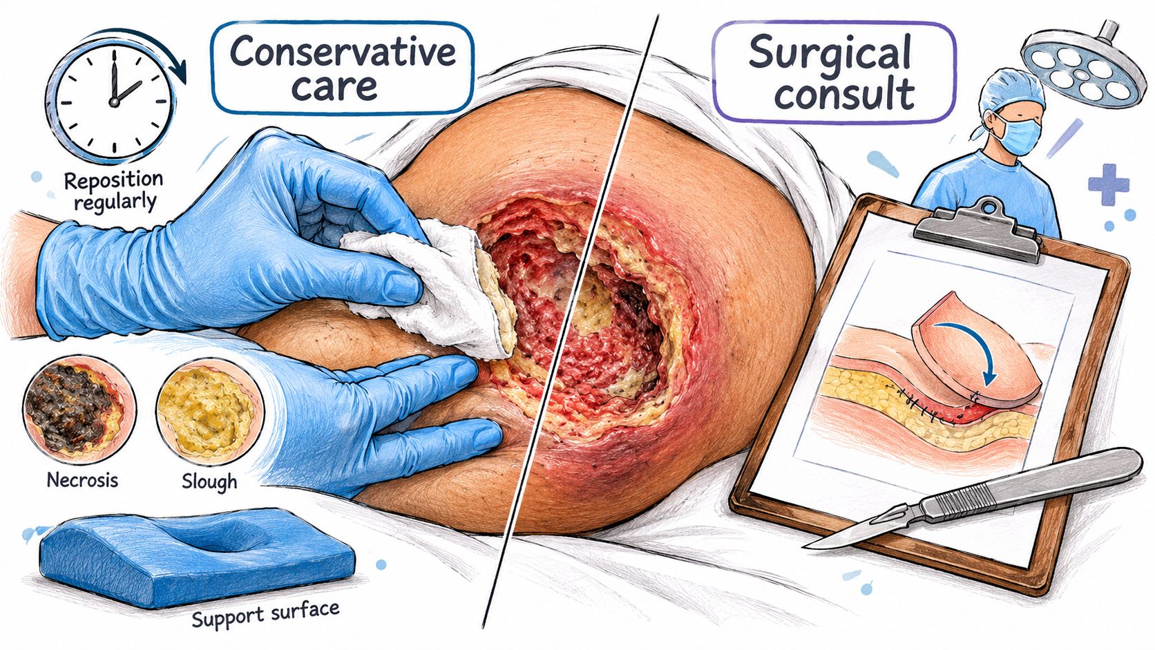
The choice is a range, not a single operation. This diagram separates conservative care from surgical review.
| Option | Typical use | Main limit |
|---|---|---|
| Debridement | Removing necrotic or infected tissue to expose a clean wound bed | Often repeated; does not close the wound on its own |
| Negative-pressure wound therapy | Healing support for stage 3 and 4 injuries under recent international guidance | Needs a sealed dressing and regular monitoring |
| Direct closure | Small, well-defined defects with enough mobile tissue to bring edges together | Limited to wounds where edges can meet without tension |
| Grafting | Clean, well-perfused bed that needs coverage | Depends on a healthy bed; poorly perfused tissue may not take the graft |
| Flap reconstruction | Deep wounds needing well-vascularized tissue over exposed bone or tendon | Major surgery; dehiscence is common and postoperative offloading is required |
Evidence has not established one surgical technique as superior, and wound dehiscence is common after repair. Recent international guidance also recommends negative-pressure wound therapy as a healing-support option for stage 3 and 4 injuries, so surgery versus dressings is a false binary.
Who reconstruction is not for
Some patients cannot safely undergo reconstruction. Severe comorbidities, poor nutrition, impaired perfusion, limited ability to maintain postoperative offloading, or goals focused on comfort can make a major operation a poor trade. A patient with a healing hip fracture, low protein stores and no reliable caregiver for postoperative positioning is not a good candidate for a flap, even if the wound looks dramatic.
The decision pathway connects findings to action. Persistent undermining or suspected deep infection may trigger imaging or bone evaluation. Spreading infection or systemic signs needs urgent action. Vascular status, nutritional repletion and glycemic control affect whether a patient is a reasonable candidate, and reconstruction requires a firm commitment to postoperative pressure relief, which is the main predictor of whether a repair holds.
The four-step pathway
Our surgical wound evaluation follows a four-step escalation pathway: begin with conservative care, read the signal when progress stops, get a virtual consultation for surgical judgment, and move straight to care when intervention is appropriate.
After a wound heals, the team should keep monitoring the closed site, maintain off-loading, and review early if the same area begins to break down. Recurrence is common when pressure returns to the same bony prominence.
The correction to hold onto: a stage 4 diagnosis opens a discussion about the right level of care. It does not schedule an operation.
Complications and Long-Term Outlook for Stage 4
A small opening can conceal substantial tissue loss, so surface area alone does not predict the course of a stage 4 pressure injury. The injury's depth and the structures involved matter, as do the person's overall health and ability to maintain pressure relief.
The figures below describe pressure injuries across patient groups and care settings. They provide context for the wider burden, not a forecast for one person.
| Measure | Reported figure | How to read it |
|---|---|---|
| Pressure ulcer prevalence among hospitalized patients (meta-analysis) | 12.8% | About 13 in every 100 hospitalized patients in the analyzed data had a pressure ulcer. |
| Estimated hospital-acquired pressure injury incidence | 8.4% | About 8 in every 100 patients developed a pressure injury during hospitalization, according to the estimate. |
| Acute-care prevalence estimates | 6% to 18.5% | Across acute-care reports, estimates ranged from about 6 to 19 patients in every 100. |
| Mortality in older patients with pressure injury within 21 months | 3.6-fold higher than comparable patients without | The comparison shows higher mortality among older patients with pressure injuries; it does not establish that the wound alone caused the difference. |
What the numbers do and do not prove
A pressure injury can occur alongside immobility, frailty, neurological disease, malnutrition, infection and other serious conditions. The reported mortality comparison does not show that a stage 4 wound alone causes death. It does show why clinicians consider the person's overall health, not just the wound.
Over time, complications can include deep infection, recurrence after healing, wound breakdown after surgery, and worsening undermining or tunnels. Undermining is tissue loss beneath the wound edge; a tunnel is a narrow channel extending from the wound. A tunnel that grows at the same clock position, new foul-smelling drainage, or increasing pain warrants reassessment.
Healing often takes time. Compare measurements across visits rather than judging progress from a single reading. Pressure relief, nutrition and treatment of underlying illness all affect the outlook.
Next Steps for a Stage 4 Wound
Bring four questions to the next visit.
- Has bone involvement, an abscess or necrosis been ruled in or out? Ask what the infection plan is, including the planned antibiotic duration and the source-control steps.
- Are the basics fully in place? Confirm pressure redistribution on a set schedule, a suitable support surface, control of moisture and friction, and a nutrition plan that someone is checking.
- When is the next formal reassessment, and how do the latest measurements compare with the baseline? Use the log from earlier visits so the team can see whether the wound is contracting.
- If the wound stalls or shows necrosis, tunneling, exposed deep structures or recurrent infection, ask for a specialist surgical wound evaluation. The review may lead to debridement, negative-pressure therapy, reconstruction or coordinated conservative care. Your current team should stay involved throughout.
At CWS Wound Services, review starts virtually: we look at the history, wound, imaging and treatment response, then a surgical wound care nurse validates the wound in person to confirm what was seen. Our escalation pathway is designed to move from conservative care to specialty judgment without replacing your current team. You can reach us at 2112 F Street, NW Suite 804, Washington, DC 20037, or by phone at 202-861-2971.
A specialist evaluation does not automatically lead to surgery. It gives you and your family a structured answer about what the wound needs next.
---
Wound Services evaluates complex, stalled and surgically appropriate pressure injuries, including stage 4 wounds, through virtual or in-person specialty review and surgical care at its own ambulatory surgical center when appropriate. If you're weighing whether a wound needs a different level of care, our team can review the history, measurements and images and send recommendations back to your current providers. Visit Comprehensive Wound Services to start an evaluation.